Tuesday, December 2 | 3:10 p.m.-3:20 p.m. | SSJ18-02 | Room N226
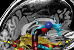
Diffusion-tensor MRI (DTI-MRI) may be a "promising additional tool" for detecting tubers and perituberal tissues in tuberous sclerosis complex (TSC) patients younger than 3 years old.Researchers from the University of California, Los Angeles (UCLA) concluded that DTI-MRI can find epileptogenic tubers, which may lead to treatment to control seizures and enhance cognitive outcomes.
Tuberous sclerosis complex patients often require invasive intracranial procedures to identify epileptogenic tissue in the brain.
"In the past, we successfully detected epileptogenic foci using FDG-PET and MRI coregistration in TSC," said lead author Dr. Akira Yogi, a visiting scholar in the division of neuroradiology at UCLA's David Geffen School of Medicine. "DTI is the other modality to evaluate molecular-level changes, which suggest the biomarker of brain epileptogenesis."
The benefit to the patient is heading to surgery without having to endure an invasive presurgical evaluation, he added.
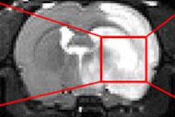
This retrospective study included 22 patients (mean age, 5.3 years) who underwent tuber resection for the treatment of epilepsy between 2004 and 2011. All patients had preoperative DTI. A total of 545 tubers were found and classified as either epileptogenic or nonepileptogenic.
Two observers manually outlined all regions of interest on an apparent diffusion coefficient (ADC) map as a reference of T2-weighted or fluid-attenuated inversion-recovery (FLAIR) imaging.
Among the findings, maximum ADC values were significantly higher in epileptogenic tubers, most notably in patients younger than 3 years.
Yogi and colleagues plan to continue their research by assessing structural and functional changes over time in epilepsy patients through functional MRI and DTI.