New MRI techniques are making it easier to diagnose cartilage damage earlier, which may result in fewer hip replacements in young patients, according to a February 18 presentation at the American Academy of Orthopaedic Surgeons (AAOS) annual meeting in San Diego.
Paul Beaulé, MD, of the Joint Replacement Institute, said he advocated reducing the incidence of hip replacement in young patients as a quality-of-life issue. While open surgery is safe and effective, the procedure can result in significant morbidity for the patient, he added.
Beaulé said new imaging techniques do more than simply capture gross morphology. By using biomarkers, a more precise portrayal of joint tissue and pathology can be provided.
 |
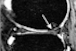
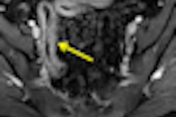
| An apparently normal lateral radiograph of the hip (above) in a patient with femoroacetabular impingement. MRI (below) of the same patient shows significant femoral head-neck offset abnormality. The arrow indicates an anterolateral head-neck junction cam impingement bump. Images courtesy of AAOS. |
 |
Panelist Kawan Rakhra, MD, from Ottawa Hospital and the University of Ottawa, explained that dGEMRIC can detect changes in glycosaminoglycan in the cartilage of a patient with arthritis. The cartilage looks normal on x-rays and standard MR images, but dGEMRIC can determine that the cartilage is biochemically abnormal.
In patients with obvious symptoms of femoroacetabular impingement (FAI) for whom radiographs and MRI scans are normal, dGEMRIC and other new imaging techniques may be useful.
The panel also noted that x-rays may be beneficial in cases of moderate to severe osteoarthritis. However, in cases with moderate or very focal chondral changes, radiographs are ineffective, and MRI should be the modality of choice. MRI allows direct visualization of hyaline cartilage and can localize and characterize fissures, thinning, and full-thickness defects, the panelists concurred.
In addition, the orthopedists extolled high-resolution MRI, which features thinner slices and no contrast agents, as a more viable option compared to MR arthrography, which can have poor sensitivity, specificity, and accuracy, as well as poor interobserver correlation.