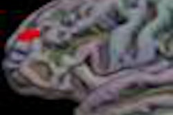
Middle-aged women who experience migraine with aura are more likely to have brain infarctlike lesions that are detectable on MRI than men or other migraine sufferers who don't have the visual phenomenon associated with their headaches, according to a study presented at last week's American Academy of Neurology (AAN) meeting in Seattle.
"This study supports the hypothesis that migraine with aura may have long-term cerebral consequences," said Anne Scher, Ph.D., assistant professor of epidemiology at the Uniformed Services University in Bethesda, MD.
The study results indicate that "a remote history of migraine with aura was associated with late-life cerebellar infarctlike lesions in women. We did not find a correlation with men," Scher said. The study included approximately 5,000 participants from the Age, Gene/Environment Susceptibility (AGES)-Reykjavik Study.
Scher noted that about 12% of adults experience migraine headaches, which appear to peak between the ages of 20 and 40 years. Migraines are two to three times more prevalent in women. About one-third of migraines are accompanied by aura -- 99% of the time the aura is described as visual, but it can also be accompanied by sensory, aphasic, and motor components.
The Reykjavik Study of men and women living in the Icelandic capital enrolled 5,764 participants, approximately 58% of whom were women; MRI scans were available for 5,003 of the subjects. The final sample from the study included 4,689 individuals, 57% of whom were women.
Participants were initially examined in the Reykjavik Study at an average age of 51 years (range, 33-65). Subjects reporting headaches once or more per month were asked about migraine symptoms including nausea, unilateral location, photophobia, visual disturbance during or just before headache (visual aura), and numbness on one side during or just before headache (sensory aura).
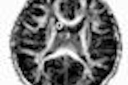
MRI exams were performed as part of the AGES-Reykjavik Study in 2002-2006. The presence of infarctlike lesions (cortical, subcortical, cerebellar, and total) was measured by trained readers blinded to migraine status. A comprehensive cardiovascular risk assessment was performed at both examinations.
Overall, the risk of having any form of late-life brain infarctlike lesion was approximately 40% greater for individuals who experienced migraine with aura in midlife, when compared with those who had migraines without aura, a history of nonmigraine headache, or people without headache. The difference was statistically significant (p = 0.005).
Scher said there was a 60% increased risk of having late-life cerebral infarcts if participants reported having migraine with aura in their lifetimes. She said there was not a significant finding for infarcts in the cortical and subcortical areas. Migraine without aura did not significantly differ from patients with nonmigraine headaches or from normal subjects.
However, when Scher and colleagues analyzed the migraine cases by sex, they found no difference in infarctlike lesions between men and normals, but a significant 90% increase in the likelihood of having the lesions among women.
By Edward Susman
AuntMinnie.com contributing writer
May 5, 2009
Related Reading
Radiofrequency ablation promising for refractory chronic cluster headache, May 5, 2009
Patent foramen ovale closure relieves migraine in patients with MRI lesions, March 6, 2009
High iron accumulation seen in the migraine brain, February 19, 2009
Migraine with aura may be linked with patent foramen ovale, November 28, 2007
Brain differences detected in migraine sufferers, November 20, 2007
Copyright © 2009 AuntMinnie.com