The clinical features of acute myocarditis can be deceptively benign: fatigue, palpitations, weakness, fever, and/or angina. But if underestimated, myocarditis can ramp up to become a chronic disease with dilated cardiomyopathy, or even mimic acute myocardial infarction. As a result, accurate assessment is key and European researchers are working with MRI to determine if it is the ultimate modality for diagnosing this disease.
First, Dr. Olaf Rieker and colleagues from Johannes Gutenberg University in Mainz, Germany, evaluated the imaging features of acute myocarditis on ECG-gated breathhold MR. Rieker presented the group’s results at the 2002 RSNA in Chicago.
For this study, 15 patients with suspected myocarditis were imaged with either a 1.5-tesla Magnetom Vision or Magnetom Sonata scanner (Siemens Medical Solutions, Erlangen, Germany). ECG-gated breathold T2-weighted images with fat suppression (TIRM sequence) and without fat suppression (HASTE sequence) were acquired in three standard views. In addition, T1-weighted imaging with FLASH was performed 10 minutes after the IV administration of Gd-DPTA. Finally, cine MRI was performed.
ECG, echocardiography, creatine kinase, troponin T and serological tests were available for all the patients, Rieker said. The MR findings were retrospectively compared to the discharge diagnoses.
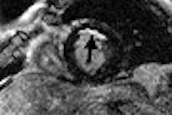
According to the results, a final diagnosis of acuyte myocarditis was established with MR in five patients. The signal intensity of the myocardium in the T2-weighted images (with and without fat suppression) of these patients was elevated. Myocardial edema was more conspicuous using the fat suppression sequence. The TIRM images could not be properly evaluated because of artifacts, Rieker explained. Finally, delayed enhancement was found in 4 out of 5 myocarditis patients.
Rieker’s group updated these results in the December 2002 issue of a German imaging journal. In this version of the study, 21 patients with suspected myocarditis underwent the same MR protocol as stated above. Acute myocarditis was diagnosed in nine patients and cardiac sarcoidosis in two patients, with late enhancement of Gd-DPTA occurring in all cases (RoFo. Fortschritte auf dem Gebiete der Rontgenstrahlen und der neuen bildgebenden Verfahren, December 2002, Vol. 174:12, pp. 1530-1536).
"MRI has the potential to show the location and extent of acute myocarditis," Rieker concluded in his RSNA talk. "MRI might be used to guide diagnostic myocardial biopsy," which is currently considered the gold standard for pinpointing myocarditis.
Myocarditis and skeletal muscle
A team of French radiologists and cardiologists went a step further by seeing if MRI would reveal acute myocarditis, as well any accompanying skeletal muscle involvement.
"Previous studies have demonstrated that either T2-weighted or Gd-enhanced T1-weighted MR sequences are helpful to diagnosing acute myocarditis, mostly by demonstrating an increased cardiac signal relative to the skeletal muscle," wrote lead author Dr. Jean-Pierre Laissy, Ph.D., from the department of radiology at Hôpital Bichat in Paris. "Yet these studies have not studied perfusion using MRI in the context of myocarditis. In addition, there may be skeletal muscle involvement in these viral infectious processes" (Chest, November 2002, Vol.122:5, pp.1638-1648).
The study population consisted of 20 patients with suspected acute myocarditis and seven healthy control subjects. MR imaging was performed on a 1-tesla Magnetom scanner (Siemens Medical Solutions, Erlangen, Germany) using ECG-gated, T2-weighted axial sequences and ECG-gated, T1-weighted sequences in the axial plane and in the short-axis view, both with and without gadolinium administration. Serial multisection, breathhold, T1-weighted turboFLASH images also were acquired. Finally, cine MRI was performed in 13 patients.
Two trained readers reached a consensus interpretation of the images. "Myocardial abnormalities included areas of high signal on T2-weighted images and on Gd-enhanced spin-echo-weighted images, judged as focal or diffuse in distribution," the authors explained.
Based on the results, all patients displayed myocardial abnormalities on subtracted T1-weighted MR that were either focal (all patients examined within seven days of onset) or diffuse (all patients examined after seven days).
Quantitatively, myocardial enhancement averaged 56% on spin-echo, T1-weighted images in the patient group. Enhancement of nodular foci was 76%. The cine-MRI showed segmental left ventricular motion abnormalities in 62% of the patients. Serial, dynamic turboFLASH images allowed for the examination of paraspinal muscle enhancement, which was observed in 95% of the patients in early- and late-stage myocarditis.
The group concluded that the MR sequences with contrast were the most useful for qualitative assessment of myocarditis. Cine MRI helped study the severity of the disease, but could not be relied upon for diagnosis. T2-weighted images turned in poor results, mainly because it did not adequately demonstrate edematous myocardial involvement.
Dynamic studies with turboFLASH revealed different enhancement patterns in the two stages of the disease, and abnormal peripheral skeletal enhancement, the author stated. These results are noteworthy because the disease may extend to the skeletal muscles.
MRI’s noninvasiveness gives it an advantage over the gold standard of endomyocardial biopsy, the group found. In addition, enodomyocardial biopsy lacks sensitivity (50%-63%) to focal disease. MRI may also go one better than 67Ga scintigraphy because the specificity of that modality is limited -- the radiotracer attaches itself to any myocardial necrosis regardless of whether it is a consequence of myocardial infarction.
"Qualitatively, contrast-enhanced T1-weighted subtracted images had 100% sensitivity and specificity for myocardial involvement," they wrote. "Post-contrast T1-weighted images were able to discriminate the early phase (nodular enhancement) from the later phase of myocarditis (diffuse enhancement)...our results (are) consistent with the hypothesis that acute myocarditis may progress from a primarily focal to a diffuse process within approximately one week."
By Shalmali PalAuntMinnie.com staff writer
March 3, 2003
Related Reading
MRI better than SPECT for predicting myocardial viability soon after MI, February 19, 2003
Cardiac MRI outdoes SPECT in spotting microinfarcts, February 14, 2003
Cardiac MRI may improve triage of patients with chest pain, February 4, 2003
Adrenergic Nervous System of the Heart, April 3, 2002
Copyright © 2003 AuntMinnie.com


