Users may rave about the effects of ecstasy, but the popular club drug could damage the brain's serotonin axons, according to Dutch researchers writing in the latest issue of Radiology.
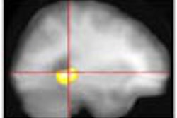
In a study that combined diffusion and perfusion MR imaging to evaluate Ecstasy-induced changes in the brain, the researchers found significantly higher diffusion coefficient values and relative cerebral volume (rCBV) maps in the globus pallidus of ecstasy users compared with nonusers (Radiology, September 2001, Vol. 220: 3, pp. 611-617).
Ecstasy, or MDMA (3,4-methylenedioxymethamphetamine) produces a rapid release of serotonin in the brain that is responsible for the drug's euphoric effects. However, MDMA is known to have toxic effects on brain serotonin neurons in long-term users, based on previous studies that show reductions in various markers unique to serotonin axons, according to the study authors, Dr. Liesbeth Reneman, Dr. Charles Majoie, and Dr. Gerard den Heeten from the Academic Medical Center in Amsterdam.
Less well known are the functional consequences of MDMA-induced serotonoergic damage. Here, brain microcirculation is an interesting starting point, since there is substantial evidence that serotonin, a vasoconstrictor, is involved in the regulation of brain microcirculation.
"It is of interest to note that findings in several case reports have linked the abuse of MDMA to the occurrence of cerebrovascular accidents, as a result of MDMA-induced effects on brain serotonin concentrations," the authors wrote.
Diffusion-weighted imaging is a promising approach for evaluating tissue changes in degenerating brain and nerve matter, because it enables the quantitative measurement of diffusional motion of water molecules in tissue, especially axons. Cellular structures, such as highly organized myelinated axons in white matter, restrict the molecular motion of water, reducing the apparent diffusion coefficient (ADC) compared to bulk water, the authors wrote. Meanwhile, dynamic contrast-enhanced perfusion-weighted MR has enabled the study of brain vasculature by measuring the rCBV.
The study compared 8 Ecstasy users (7 men, 1 woman, mean age 27.6 years) with 6 Ecstasy nonusers (3 men, 3 women, mean age 22.3 years), who had also used other drugs. The healthy subjects had no history of psychiatric illness. They were group-matched by age and sex. They agreed to abstain from psychoactive drug use for at least 3 weeks before the study, and abstinence was verified by enzyme-multiplied immunoassay urine tests, the authors wrote.
Diffusion and perfusion MR imaging were performed with a 1.5-tesla Magnetom Vision scanner (Siemens, Erlangen, Germany) following the acquisition of transverse T1- and T2-weighted standard spin-echo sequences.
Echo-planar imaging was used to obtain diffusion images (700/118, 1 signal acquired, 23-cm FOV, 96 x 200 matrix), with diffusion gradients applied independently on each axis of the magnet. Beginning with a baseline image using minimum diffusion weighting, 9 diffusion-weighted images were obtained along each axis, using progressively higher b values to describe the diffusion sensitivity of each sequence.
"Although signal sensitivity in the diffusion-weighted imaging is affected by T1 and T2, the ADC map is not. It is obtained by calculating the logarithmic ratio of the signal intensity at each pixel...." the authors wrote.
They also acquired echo-planar T2-weighted dynamic images (0.8/54 1 signal, 23-cm FOV, 128 x 128 matrix). The images were obtained in 12 transverse sections at 1.2-sec. intervals for 64 seconds immediately after bolus injection of 0.1 mmol gadopentetate dimoglumine per kg of body weight.
Images were reconstructed using software from the Massachusetts General Hospital's nuclear magnetic resonance center in Charlestown, MA. Regions of interest in the left and right frontal cortices, the occipital cortex, white matter, putamen, and globus pallidus were evaluated by a radiologist who was blinded to the subjects' histories. Mean signal intensities were measured in each ADC and rCBV map, and compared to a white-matter reference standard.
The researchers administered the Dutch Adult Reading Test (DART-IQ) to obtain an estimate of verbal intelligence, and assessed differences between the two study groups using the Mann-Whitney-Wilcoxon test.
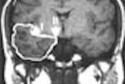
According to the results, conventional T1- and T2-weighted images showed no edematous changes in the brains of Ecstasy users and nonusers, and there were no significant differences between left and right ADC and rCBV ratios.
Mean ADC values were higher overall in Ecstasy users compared with nonusers, but statistically significant only in the globus pallidus (0.65 x 10⁻⁵ cm2/sec in nonusers, vs. 0.84 x 10⁻⁵ cm2/sec in Ecstasy users). rCBV values followed a similar pattern, with higher values reaching statistical significance only in the globus pallidus (mean, 1.01 in nonusers, vs. mean, 1.22 in users).
"No significant correlations were observed between ADC values and extent of previous Ecstasy use," the authors wrote. "Age, sex, and DART-IQ test results were not significantly associated with rCBV ratios or ADC values. However, a significant association was observed between extent of previous Ecstasy use and rCBV ratio in the globus pallidus...," they noted.
Because experimental models have shown that the axonal cell membrane is sufficient to account for most of the restriction of water diffusion in white matter, any process that disturbs the integrity of the axon would logically change the diffusion of water in this tissue, the authors wrote.
Thus, higher ADC values in the globus pallidus of Ecstasy users may be due to axonal injury or vasogenic edema. Previous studies have shown serotonergic axonal loss of 80%-90% in various brain regions of animals treated with MDMA, and the damage has also been demonstrated anatomically by the use of cytochemical methods for visualizing axons that contain serotonin, according to the authors.
As for the higher rCBV values in ecstasy users, significant evidence points to a strong vasoconstrictor role of serotonin in the control of brain microcirculation, they wrote.
"It is thought that, because of reduced serotonin content, vasodilation occurs due to the removal of serotonergic constrictor effects," the researchers said. "The increases in rCBV ratios in the globus pallidus of Ecstasy users in this study may result from a similar mechanism."
Still, the sample size was small, and there is a chance that people with higher pallidal rCBV ratios and ADC values are predisposed to use Ecstasy, the authors noted. Ecstasy users also reported higher methamphetamine use than non-Ecstasy users, and while that drug's effects were likely insignificant, they could not be completely ruled out.
The authors concluded that Ecstasy use is suggestive of changes in rCBV ratios and ADC values in the globus pallidus.
"These findings are consistent with findings of serotonergic axonal loss and serotonin depletion in animals treated with MDMA, data in humans from other published reports, and findings of cerebrovascular accidents in medical histories of Ecstasy users," they wrote. "Our data indicate that MR imaging may be a valuable tool in the investigation of the consequences of MDMA use in brain tissue and the microvasculature."
By Eric BarnesAuntMinnie.com staff writer
September 18, 2001
Copyright © 2001 AuntMinnie.com


