A group in Australia has described a rare finding discovered during a prostate cancer restaging PET/CT scan – a pulmonary tumor embolism, according to a case published September 19 in Radiology Case Reports.
The case underscores the importance of high-quality prostate-specific membrane antigen (PSMA) radiotracers for detecting incidental findings, suggested lead author Jared Tan, of the University of Melbourne in Australia, and colleagues.
"With increased diagnostic power comes the responsibility to detect rare, previously disregarded lesions," the group wrote.
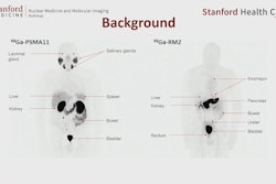
In this case, a 75-year-old woman presented with recent dyspnea and left-sided chest pain. Her history included a right nephrectomy for clear renal cell carcinoma (RCC) with renal vein involvement four years earlier. She had been treated with a tyrosine kinase inhibitor and was in remission. Hybrid contrast-enhanced CT (with iodinated contrast bolus timed to maximize pulmonary arterial visualization) and F-18 DCFPyL PSMA-PET images. Contrast CT images (A, B) show an eccentric filling defect in the superior lingula segmental artery of the left lung upper lobe (red arrow). This is concordant with moderately intense PSMA radiotracer expression (black arrows on axial PET-only images, C, and on whole-body maximum intensity projection image, E). Fused PET and contrast-enhanced CT axial image (D) also confirm the lesion (red arrow). No other site of PSMA expressing metastasis is evident on whole-body imaging (E).Radiology Case Reports
Given her new symptoms, the clinicians opted to restage with hybrid F-18 DCFPyL (Pylarify) PSMA-PET and contrast-enhanced CT. The imaging demonstrated abnormal PSMA tracer uptake in the superior lingula segment of the left upper lobe of the lung. This was confirmed on high-resolution fused PET/CT images to be an eccentric filling defect in the lingula segmental pulmonary artery.
"Given the PSMA expression, this was interpreted as a new metastatic tumor thrombus. No other site of PSMA expressing metastatic disease was evident," the group wrote.
The patient was started again on pazopanib tyrosine kinase inhibitor therapy, which led to regression of the lesion on subsequent CT imaging, with the patient's chest pain and dyspnea gradually improving, they wrote.
"To our knowledge, this is the only case report describing a [pulmonary tumor embolism] in a patient with RCC detected by PSMA-PET/CT," the doctors wrote.
Despite the name, expression of PSMA is not limited to prostate cancer, the authors noted. RCC subtypes (predominantly clear cell) have also been shown to overexpress PSMA. Ultimately, the development of novel PSMA-targeting agents in PET/CT has empowered imaging specialists and oncologists with profound diagnostic capability, they wrote.
"But in doing so, it opens the doors to a multitude of previously unconsidered diagnoses," the authors concluded.
The full article is available here.