A novel PET radiotracer that visualizes synaptic density outperformed FDG-PET in detecting brain changes associated with behavioral variant frontotemporal dementia, a Yale University study has found.
The study is the first to directly compare F-18 SynVesT-1 – which targets a protein on synapses called synaptic vesicle glycoprotein 2A (SV2A) -- and F-18 FDG-PET within the same individuals with the disease, noted lead author Arman Fesharaki-Zadeh, MD, and colleagues.
"The ideal biomarker for [behavioral variant frontotemporal dementia] is still missing, highlighting the ongoing need for more robust and disease-specific neuroimaging biomarkers," the group wrote. The study was published April 9 in Alzheimer’s & Dementia.
Behavioral variant frontotemporal dementia (bvFTD) is the most common form of FTD, characterized by progressive, insidious changes in personality, empathy, and social behavior due to degeneration in the brain's frontal and temporal lobes. Nearly half of cases are initially misattributed to psychiatric conditions, resulting in an average diagnostic delay of five to six years, the authors noted.
F-18 FDG-PET is the established imaging standard for diagnosing bvFTD, yet it is not specific and reflects overall glucose uptake rather than directly measuring neuronal or synaptic loss, the group added. To further validate F-18 SynVesT-1 as an alternative, the researchers conducted a head-to-head comparison between the tracers.
Between December 2021 and September 2024, the group recruited 20 participants, including 10 with bvFTD (mean age 70.6; six men, four women) and 10 healthy controls (mean age 67.6; four men, six women), with all subjects undergoing both scans.
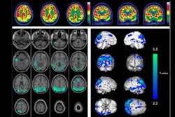
 (A) Averaged parametric images of F-18 SynVesT-1 BPND values and (B) F-18 FDG SUVR (60–90 minutes) values overlaid on anatomical (T1) images in MNI space show group differences between behavioral variant frontotemporal dementia (top) and healthy controls (middle). The color scale represents (A) BPND and (B) SUVR values. On the bottom, average z-score maps were generated to visualize regional reductions in F-18 SynVesT-1 BPND and F-18 FDG SUVR in bvFTD relative to healthy controls. For each voxel, z-scores were calculated as z = (bvFTD−meanHC )/SDHC. Negative z-scores indicate lower synaptic density or glucose metabolism in the bvFTD compared with controls in the regions. BPND, non-displaceable binding potential; bvFTD, behavioral variant frontotemporal dementia; F-18 FDG, F-18 fluorodeoxyglucose; MNI, Montreal Neurological Institute; SUVR, standardized uptake value ratio. Alzheimer’s & Dementia
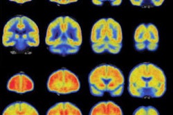
In addition, SV2A reductions also extended to secondary regions including the striatum (–32%), hippocampus (–29%), and thalamus (–23%); only SV2A binding correlated significantly with executive function, the researchers reported.
“We found that lower synaptic density was more pronounced than hypometabolism across all primary regions, and only SV2A binding correlated with clinical measures of executive function,” the group wrote.
As this was the first study to provide in vivo evidence comparing the two tracers, larger prospective studies with genetic profiling and pathological confirmation are needed, the researchers noted. They also highlighted that SV2A quantification currently requires labor-intensive methods not yet in routine use.
"As precision diagnostics and targeted interventions evolve, SV2A PET may offer a robust, sensitive biomarker for disease staging, treatment monitoring, and clinical trial endpoints in bvFTD," the group concluded.
The full study is available here.