NEW ORLEANS – A group from Shandong First Medical University has won this year’s Image of the Year award at the Society of Nuclear Medicine and Molecular Imaging annual meeting for the development of a new immuno-PET tracer.
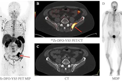
The image illustrates that a first-in-class peptide-based PET tracer called F-18 AlF-NOTA-PCP2 can more effectively measure programmed death-ligand 1 (PD-L1) expression in patients with head and neck cancers than the conventional F-18 FDG tracer.
“Our study demonstrates the feasibility of F-18 AlF-NOTA-PCP2 for real-time PD-L1 imaging in [head and neck carcinoma], paving the way for optimized treatment strategies,” noted first author Yong Wang, MD, in a news release.
PD-L1 is a highly expressed protein in head and neck squamous cell carcinoma (HNSCC). While some studies have reported that F-18 FDG-PET can predict tumor PD-L1 expression, others have presented contradictory findings, the researchers noted.
“To address this limitation, we developed F-18 AlF-NOTA-PCP2, a novel PD-L1-targeted peptide-based tracer designed to enhance hydrophilicity and achieve superior tumor-to-background ratios," added co-author Man Hu, MD. "Compared with [gallium-68] analogs, the F-18 AlF tracer can be produced with a high-yield dosage."
First, the group assess the PD-L1 binding specificity of F-18 AlF-NOTA-PCP2 in preclinical imaging in mice with head and neck cancer xenografts. These studies showed that F-18 AlF-NOTA-PCP2 binds to PD-L1 with high affinity and specifically targets PD-L1-positive tumors.
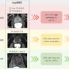
Next, in a clinical study in 24 patients with HNSCC, participants underwent F-18 AlF-NOTA-PCP2-PET/CT imaging, with 16 also receiving paired F-18 FDG-PET scans for comparison. Researchers analyzed tracer uptake, safety, biodistribution, and the correlation between imaging results and PD-L1 expression confirmed by immunohistochemistry. PD-L1-targeting molecular imaging in head and neck carcinoma: A head-to-head comparison of F-18 AlF-NOTA-PCP2-PET and F-18 FDG-PET/CT.Yong Wang, MD, and SNMMI.
According to the findings, tracer uptake strongly correlated with PD-L1 expression measured by immunohistochemistry, confirming its ability to noninvasively detect PD-L1 in vivo. In addition, a direct comparison with F-18 FDG PET revealed only a moderate correlation, highlighting the limited ability of F-18 FDG to predict PD-L1 expression, the authors noted.
“Combining FDG-PET with immuno-PET tracers like F-18 AlF-NOTA-PCP2 could offer a more comprehensive understanding of tumor biology, improving patient selection and informing tailored immunotherapy strategies,” Wang said.
According to Heather Jacene, MD, SNMMI Scientific Program Committee chair, the introduction of a targeted, specific tracer for PD-L1 expression could be transformative for molecular imaging and nuclear medicine.
Pending further studies and regulatory approval, 18F-AlF-NOTA-PCP2 could become available for clinical use in the next two to three years, the authors noted.
“This advancement has the potential to significantly enhance the precision of imaging in oncology, enabling clinicians to more accurately track immune-related biomarkers and make real-time adjustments to treatment strategies,” she said.
Each year, SNMMI chooses an image that best exemplifies the most promising advances in the field of nuclear medicine and molecular imaging. This year, the SNMMI Image of the Year was chosen from more than 1,300 abstracts submitted for the meeting.