Researchers have developed a new PET imaging system that significantly increases spatial resolution and could lead to improved detection of pathology associated with Alzheimer's disease, according to a study presented June 13 at the annual meeting of the Society of Nuclear Medicine and Molecular Imaging (SNMMI).
A French and U.S. team of computer engineering and radiology investigators combined FDG-PET with a real-time infrared camera that tracks motion and produced superresolution scans with visibly better spatial resolution compared with static acquisitions. The system could overcome PET limitations related to patient movement when performing brain imaging, according to the researchers.
"We show it's actually possible to harness that usually undesired motion to enhance the spatial resolution in brain PET using a technique called superresolution," said lead author Yanis Chemli of the Gordon Center for Medical Imaging in Boston.
Patient movement significantly reduces spatial resolution in PET and makes it more difficult to identify pathological biomarkers in the brain associated with disease, such as tau protein in Alzheimer's disease. Superresolution imaging technology typically refers to reconstruction techniques that synchronize low-resolution images or sequences of the same object to form high-resolution images. The technique has been applied in medical imaging with varying results.
In this study, the researchers sought to develop and evaluate a superresolution estimation framework for brain PET that precisely and continuously measures object shifts by taking advantage of a high-resolution infrared motion-tracking camera.
Studies with Hoffman 3D brain phantoms and nonhuman primates (rhesus monkey) were performed on a PET/CT scanner (Discovery MI, GE Healthcare). The external motion tracking camera (Polaris Vega, NDI), which yields a tracking accuracy up to 0.12 mm, was integrated with the scanner through a custom-built interface.
Brain phantoms filled with F-18 FDG were scanned for 15 minutes in list mode while undergoing continuous rotation and translation movements. The researchers simulated head movement by moving the bed, shifting and rotating the subject, and by moving the reconstruction matrix. Similarly, a rhesus monkey was injected with F-18 FDG and scanned for 15 minutes in list mode with continuous head motion induced manually. Reference static PET acquisitions were performed without inducing movement.
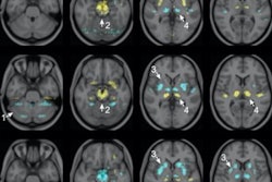
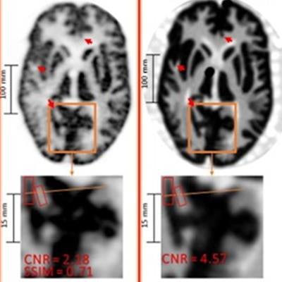
 Result of the Hoffman brain phantom study. Top row: same PET slice reconstructed with 2-mm static ordered subset expectation maximization (OSEM), (B) 1-mm static OSEM, proposed superresolution (SR) method, and corresponding CT slice (note that the CT image can be treated as a high-resolution reference). (Middle row) Zoom on region of interest for corresponding images. (Bottom row) Line profiles for corresponding data. Image courtesy of Yanis Chemli.
For both the phantom and monkey studies, the combined superresolution system resulted in PET images with visibly increased spatial resolution, compared with static acquisitions reconstructed using 1- or 2-mm voxels. The new images provided improved visualization of small cortical and subcortical brain structures, according to the researchers.
Overall, the new method achieved better noise control than static reconstructions with the same voxel size due to the interpolation of continuous motion transformations during image estimation. Line profiles confirmed improved spatial resolution for the superresolution images in both the phantom and animal studies.
"By harnessing motion and using a high-resolution tracking device, we can actually achieve superresolution and reconstruct the images beyond the scanner's intrinsic resolution," Chemli said.
"While we only tested the superresolution technique in preclinical studies, we are currently working on extending it to human subjects. In the future, we hope to better image tau deposition in early stages of Alzheimer's disease with current clinical scanners," he concluded.