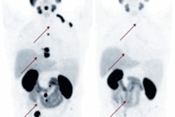
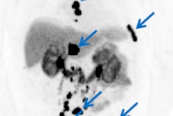
A molecular biomarker used in PET imaging can detect fast-growing prostate cancer and distinguish it from benign lesions, according to a study published in the Journal of Nuclear Medicine.
Researchers from Johns Hopkins University in Baltimore tracked 13 patients with primary prostate cancer who were imaged with F-18 DCFBC prior to scheduled prostatectomy. Twelve of the patients also had pelvic prostate MR imaging (J Nuc Med, July 1, 2015, Vol. 56:7, pp. 1003-1010).
Lead author Dr. Steven Rowe and colleagues found that, although the sensitivity of F-18 DCFBC PET imaging for primary prostate cancer was lower than MRI, it was able to detect the more clinically significant high-grade and larger-volume tumors with higher specificity than MRI.