Wednesday, December 4 | 3:00 p.m.-3:10 p.m. | SSM17-01 | Room S505AB
While whole-body-MRI is a feasible method for staging colon cancer patients, the modality does not quite reach the accuracy of FDG-PET/CT, according to a new study by Italian researchers.Researchers from the University of Rome "Tor Vergata" evaluated 40 consecutive patients with previously diagnosed colon cancer. All patients underwent 3-tesla whole-body MRI (Achieva, Philips Healthcare) and whole-body FDG-PET/CT (Discovery ST 64, GE Healthcare) to stage lymph nodes and distant metastases after resection of the primary tumor.
Regional lymph node involvement was correctly diagnosed as node-positive in 30 (75%) of 40 patients at whole-body-MRI and in 36 (90%) of 40 patients at PET/CT. Overall staging for distant metastases was accurate in 34 patients (85%) using whole-body MRI, compared with 36 patients (90%) imaged by PET/CT.
Four patients were overstaged regarding lymph node status with whole-body MRI, compared with only one patient with PET/CT. Six patients were understaged with whole-body MRI, compared with two patients with PET/CT.
For distant metastases, two patients were overstaged with PET/CT and three were understaged; with whole-body MRI, one patient was overstaged and four patients were understaged.
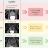
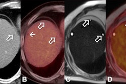
The researchers also suggested that whole-body MRI fused with contrast-enhanced T1-weighted diffusion-weighted imaging (DWI) could be an alternative to conventional whole-body methods for detecting primary tumor and distant metastases.
By improving the diagnostic process, they hope to provide a greater amount of information that can help physicians and patients choose the most appropriate therapy.