When we hear the term occupational hazard, most of us think of jobs that seem inherently dangerous: the window washer who hangs high atop a building, or the firefighter who risks his life for others. But even professions that aren't considered inherently risky can sometimes be dangerous. Under certain circumstances, radiology is one of these.
This article is intended to trace the development and pathophysiology of work-related injuries, and make radiology practitioners more aware of the occupational hazards of their jobs. I also will discuss prevention and treatment. Hopefully, my own experience will help others avoid what I've endured.
My own realization of radiology's occupational hazards was a tragic surprise. I am a board-certified diagnostic radiologist, fellowship-trained in angiography and interventional radiology. I joined a teaching hospital group and much of my work was centered in the special studies suite. It was normal to spend 8-12 hours a day, two to five or more days a week, wearing a lead apron.
Because of a ceiling-mounted monitor that did not rotate 360º, I found myself contorting my body while doing cases. My upper body would be pulled down by the weight of the apron and a face shield attached to my shoulder with Velcro. Often I had to turn my head back more than 100º to see the monitor. I would remain in this position for hours at a time. Between my small body habitus and the physical stress of the cases, this position proved to be extremely damaging over time.
I developed bilateral brachial plexus stretch and traction injury, with severe and permanent nerve damage. At the time of diagnosis in 1997, I had noticeable muscle atrophy, significant muscle weakness, excruciating diffuse nerve pain, and total debilitation.
I have spent the ensuing four years battling chronic pain and continuing functional limitations. I have not been able to practice radiology since November 1996. I feel as though I have been robbed of major aspects of my life, both professional and personal.
Had I not experienced this degree of pain and debilitation, I do not think that I would have been able to empathize with any patient in a similar situation. When the injury struck, I had just become partner in my group, and was about to take over the division. I had basically arrived at the place I had worked my whole life to reach.
I experienced excruciating bilateral nerve pain from my neck to my back and down both arms and hands. It felt as though my nerves were being cut with a knife, one by one. I had intense muscle fasciculations, myalgias, severe allodynia, hyperesthesia, weakness, and secondary insomnia.
My husband had to help me in the shower, help me dress, cut my food. I couldn’t so much as turn a doorknob or pull the cord to open the blinds. I had gone from a successful, hard-working, independent professional to an invalid, a despondent mess.
Therapeutic trials and tribulations
Fortunately, I was diagnosed early and treated by Dr. Robert Schwartzman of Hahnemann Medical University in Philadelphia. Dr. Schwartzman is a recognized expert in brachial plexopathy and reflex sympathetic dystrophy (RSD). He told me that he saw between 3-5 new cases a week of similar injuries, mostly among interventional radiologists, invasive cardiologists, and dentists.
Due to the RSD, my range of motion was so limited that even small attempts at physical therapy, such as rotating my head only a few degrees, would result in a completely new set of symptoms: My pupils would dilate; I’d become vagal, flushed, and intensely hot. I developed vertigo and hypotension. My physical therapist kept smelling salts in the office for patients like me who passed out from the sympathetic stimulation.
After five months of physical therapy, Dr. Schwartzman felt it was time for my first nerve block. He sent me to his colleague, Dr. Alyssa LeBel, a neurologist and anesthesiologist, who specialized in interpleural catheter nerve blocks.
In this procedure, the interpleural catheter is inserted intercostally using local anesthesia. Using standard techniques, a very thin catheter is passed coaxially through a needle into the pleural space. Round-the-clock bupivicaine is injected every 4 hours with the patient lying in lateral decubitus, Trendelenburg position. The goal is to quiet down the abnormal firings of the autonomic nervous system by anesthetizing the stellate ganglion, thereby breaking the pain circuit. Each treatment required a 4-6-day stay in the hospital.
I eventually went through three interpleural catheter nerve blocks, and went on to make small advances in physical therapy. I also went through several pain medications, tried acupuncture and herb therapy water therapy, massage, exercise. I did Tai Chi, visited a chiropractor, and even tried biofeedback/relaxation techniques.
Unfortunately, RSD is progressive, so the symptoms began anew in my low back and both lower extremities. Just as I was finally showing improvement in upper body pain levels and function after two years of treatment, I again found myself in terrible pain, but distal to the area of initial injury.
I had to begin therapy again, this time a series of epidural steroid injections, more physical therapy, and more drugs. I am now undergoing a combination of acupuncture/trigger point injections for this lower extremity involvement.
All of the various treatments have one purpose: to provide sufficient pain relief that I can tolerate getting out of bed in the morning and facing daily activities. Unfortunately, I still have permanent nerve damage, which prevents me from doing many simple tasks. For instance, it is impossible for me to hold a book for more than a minute, as my upper arm muscles begin to fatigue and then spasm.
I have lost coordination, so I tend to lose my balance and drop things easily and often. I have regained some strength, but I get muscle fatigue and shooting nerve pains. Every physician who has treated me has said: "I think I can help you, but you’ll never get back to being who you were." That’s a bitter pill to swallow.
Like many of you, I never thought that something like this would happen to me. I never imaged that I would need a personal disability policy even though I knew it was prudent. I knew the word ergonomics and what it meant. But I ignored it and just kept on going. It is my hope that you will take a good hard look at your own work habits and realize that this could happen to you.
Beyond anecdotal evidence
Many radiologists are aware of the anecdotal dangers of the lead apron. We all know of colleagues who have developed chronic neck or low back pain, attributable to continued use of the lead apron.
Several AuntMinnie.com articles report on cases of carpal tunnel syndrome in Australian, American, and Canadian ultrasonographers.
Concern over the hazardous situation that radiologists are putting themselves in is growing. At the 2001 American Roentgen Ray Society conference in Seattle, Dr. Lynne Ruess from the Tripler Army Medical Center in Honolulu presented the results of a study that tracked the overuse injuries in four radiologists who had complained of upper extremity pain.
An occupational therapist diagnosed several problems, including carpal, bilateral, and cubital tunnel syndromes. An industrial hygienist determined that the group’s working conditions in the reading areas and offices were putting the doctors at risk, Ruess said.
In a letter to the American Journal of Neuroradiology, Dr. David Pelz discussed the Pinkerton "Hang’em High" Apron Support System (Cook/Inoray, Niagra Falls, NY), designed to alleviate back and neck pain for interventional radiologists.
Wearing a 15-lb apron can place pressures of up to 300 lbs per square inch on intraverterbal disks," wrote Pelz, who is an associate professor at the University of Western Ontario in London, Canada. "For the angiographer suffering from back pain, the ideal apron should be weightless. The [Pinkerton system] allowed me to return to performing angiography and interventional procedures five weeks after a lumbar discectomy" (AJNR, August 2000, Vol.21:7, p.1364).
Dr. John Healy from the University of California, San Diego responded to Pelz’s letter, stating "I have been able to participate in many sports with negligible low back and neck pain, but have nearly always been symptomatic with back pain and radiculopathy when performing interventional procedures" (AJNR, January 2001, Vol.21:1 p. 221).
At the 2001 Society of Cardiovascular and Interventional Radiology meeting in San Antonio, Dr. Lindsay Machan, an associate professor of radiology at the University of British Columbia, in Vancouver, discussed the results of a survey that quizzed SCVIR members about their incidence of neck and back pain. Some of the respondents said the pain had been intense enough to force them to miss work.
Evidence of injury
Let’s review the average workday of the radiologist. It might begin at the computer terminal, signing off on reports, which may mean half an hour or more of sitting at the keyboard. Let’s say you are assigned to CT that day, so you may be sitting with a mouse for hours processing and reviewing your studies.
If your department is not yet filmless, there may be huge folders of CT scans you must pick up, push around, and sort through. Maybe you have to hang and remove your films much of the day. The scenario may be similar for MRI.
If you read mammograms, think of how long you sit, hunched over a viewbox, holding a magnifying glass, sorting comparison studies. In ultrasonography, you may find yourself in physically stressful positions while scanning patients and reviewing your films.
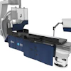
Special studies may present the most physically demanding activities. The lead is heavy and a constant habiliment. Depending on the room arrangement, monitors may be impossible to move adequately and the x-ray table may need frequent shifting. Add on the hours and strain of processing the digital films at the computer keyboard, doing your own filming, and reviewing of previous studies.
These various scenarios can lead to different injuries:
- Repetitive strain injury (RSI)
- cervical and lumbar disc problems
- carpal tunnel syndrome
- brachial plexus injury and RSD
An in-depth report on the Occupational Safety and Health Administration (OSHA) Web site describes this multitude of work-related injuries as musculoskeletal disorders (MSDs). "These MSDs develop during the performance of specific work tasks or combinations of tasks over a prolonged period of time…resulting in small and repeated tissue damage to muscles, tendons, joints, or nerve structures," the report says.
|
The OSHA Website’s excellent list of physical work
activities and ergonomic risk factors may be adapted for relevancy to the
radiologist’s day as follows: |
||
|
|
||
|
Physical Work
Activities |
Ergonomic |
Radiologist’s |
|
|
|
|
|
Exertion of considerable physical effort to complete a
motion or task |
Force Awkward/static postures Contact stress |
Moving a table/patient in GI US, biopsies, Special Studies
(SS), lead aprons |
|
|
|
|
|
Doing same motion over and over again |
Repetition Force Awkward/static postures |
US, computer terminal work, moving patients on tables,
(GI, SS), CT, and MR rooms |
|
|
|
|
|
Performing tasks that involve long reaches |
Awkward/static postures Force |
GI, US, SS, biopsies, and drainages |
|
|
|
|
|
Working surfaces too high or too low |
Awkward/static postures Force Contact stress |
Computer terminal work, CT procedures, venograms, and
other procedures |
|
|
|
|
|
Maintaining same position or posture for long periods of
time while performing tasks |
Awkward/static postures Force Contact stress |
Computer terminal work, SS, US, GI |
|
|
|
|
|
Sitting for long periods of time |
Awkward/static postures Force |
General film reading in all specialties, computer terminal
work |
|
|
|
|
|
Using hand and power procedures, tools |
Awkward/static postures Force Contact stress |
US, CT-guided SS |
|
Information courtesy of Occupational Health and Safety
Administration, U.S. Department of Labor |
||
Ergonomic risk factors
Many potential ergonomic risk factors have been sited as contributing factors to MSD, according to the National Institute for Occupational Safety and Health (NIOSH) and others. These ergonomic risk factors cause injury by imposing biomechanical stresses on the practicing radiologist in the following ways:
Ergonomic Definitions |
|
|
Force |
Tasks requiring more vigorous physical force to perform
place greater biomechanical stresses on the body. Muscles fatigue more
easily, and the musculoskeletal system in general becomes more prone to
irritation, inflammation, strains and tears. Forces may be internal, such as
tension within a muscle with movement; or external, such as moving or wearing
heavy devices. |
|
|
|
|
Repetition |
The performance of a task or movement over and over again,
for prolonged periods of time. With inadequate time for recovery, this causes
fatigue and strain. This is especially severe when a specific body part is
more involved than the rest of the body, and is especially common in the
upper extremities. |
|
|
|
|
Awkward Postures |
Performance of tasks where the position of certain parts
of the body differ significantly from the rest of the body’s neutral
position. A good example of this
would be twisting the back or torso while moving a heavy object, such as
occurs in the GI suite: moving the
monitor and/or patient table while performing the study and watching the
monitor. |
|
|
|
|
Static Postures |
When the body is kept in the same position or posture for
long periods of time, this impedes blood flow to the involved areas. As a result, the in and outflow of
nutrients and wastes of working muscles is impeded, causing increased fatigue
of the involved muscles. The muscles
then become more prone to injury. |
|
|
|
|
Contact Stress |
This is the result of occasional, repeated or continuous
contact between susceptible body parts and hard, soft, or heavy objects. The contact creates pressure over the body
part, impeding blood flow, tendon, muscle, and nerve function. Besides the example of heavy lead aprons,
another example of this is the resting of wrists on the sharp edge of a desk
while using a computer or typewriter keyboard. |
|
Information courtesy of The Repetitive Strain Injury Recovery Book, Deborah Quilter,
Walker and Co., New York, NY: 1998 |
|
MSD symptoms
In her excellent book The Repetitive Strain Injury Recovery Book (Walker and Co., New York, NY, 1998), Deborah Quilter described the evolution of MSD as starting with "a whisper and end[ing] with a scream. By the time most people realize they are injured, much damage has occurred." As my own symptoms developed over more than three years, what started as an evanescent, gnawing, focal trapezius pain ended in disability.
Much of the problem lies in the fact that those of us who work long, hectic hours often dismiss our "aches and pains." We may be aware of the evolving pain, but ignore it because we’re too busy. Some will acknowledge pain and work through it as it worsens. Others will deny or try to hide the injury for fear of losing their jobs.
Warning signs of MSD’s/RSI’s |
|
|
Pain |
Sustained or chronic, shooting, burning or aching, focal
or diffuse, experienced while working or at home |
|
|
|
|
Fatigue |
Lack of endurance in the injured area or body part |
|
|
|
|
Weakness |
in the involved area |
|
|
|
|
Coldness |
The involved body part may feel “cold”. Implies nerve
involvement or damage to blood vessel innervation |
|
|
|
|
Allodynia |
Painful sensation on skin from non-injurious stimuli, for
example, cool breeze causes pain |
|
|
|
|
Hypersensitivity, Hyperaesthesia |
Unusually increased or altered sensitivity to sensory
stimuli |
|
|
|
|
Heightened awareness |
Enhanced or excessive awareness of a body part may be an
early warning sign from your body that an injury is evolving |
|
|
|
|
Heaviness,
sluggishness, fatigue |
The involved area may feel like a “dead weight |
|
|
|
|
Difficulty
using the involved part, decreased strength or range of motion |
Stiffness, lack of control or coordination, cramping |
|
|
|
|
Tingling, numbness, clumsiness, loss of sensation, tremor,
stiffness, cramping |
|
|
|
|
|
Frequent
self-massage, avoidance of using the involved body parts, guarding |
|
|
|
|
|
Deformity,
loss of function |
|
|
|
|
|
Loss of
sleep due to pain |
|
|
|
|
|
Absence
from work or inability to perform daily activities |
|
|
Information courtesy of Repetitive Strain Injury: A Computer User’s Guide, Emil
Pascarelli, MD and Deborah Quilter, John Wiley & Sons, New York, NY: 1994 |
|
Facing reality: Taking preventive action
As physicians, most of us have worked long and hard to achieve professional success. Once attained, it's difficult to come to terms with events in our lives that might threaten that success. In addition, the modern medical practice calls for longer hours with less ancillary help and multitasking.
Professionals often deny that anything is wrong, especially if the symptoms are evanescent, as they often are in the beginning. Some will see an admission of injury as a personal failure. Others will try and self-medicate without a true diagnosis, exacerbating the injury. Some will find ways of hiding the symptoms and get through the day with subtle excuses.
So what can be done to prevent the development of job-related injuries? There are several options, but they will require the participation of multiple parties.
- Manufacturers : Bring back floor-mounted, moveable monitors. The ceiling-mounted style is probably one of the greatest contributing factors to the development of RSI/MSD in radiologists. If the ceiling-mounted monitor can't be rotated so that it's always at eye level and in front of the radiologist, injury is more likely. When new exam rooms are designed, practitioners must demand the inclusion of versatile equipment.
- Ergonomics : When planning department set-ups, good ergonomics must always be taken into account regardless of the modality. An expert consultant is worth the expense to minimize risk. Department in-service education and enforcement of good habits is crucial to any program’s success. Periodic reviews for conformity are essential.
- Professional discussion : Professional organizations, journals, and local radiology societies must increase awareness by sponsoring nationwide surveys and conferences. Collectively, this can enhance awareness on the part of manufacturers, hospitals, and among other practitioners, and is the first step to conquering the problem.
- Personally : At the first sign of a problem, it is imperative that the radiologist seek help. Although a change in work responsibilities may be the only solution, it is worth it if it prevents severe injury, debilitation, chronic pain, and disability. With limited time or exclusion from assignments, physical therapy will aid recuperation. Lastly, if arrangements cannot be made to assist recuperation, changing jobs must be a considered.
According to OSHA, work-related injuries account for one-third of all occupational injuries and illnesses that are annually reported to the Bureau of Labor Statistics. In 1997, 626,000 workdays were lost to cases of MSD. It is estimated that $15 billion to $20 billion in claims for worker’s compensation go to work-related injuries.
But these numbers pale in comparison to the effect on a person’s life, as well as the effect on his or her co-workers and loved ones. Education imparts power, and the more we are aware of this pervasive and potentially devastating problem, the more we can prevent it.
By Dr. Judith K. WeinsteinAuntMinnie.com contributing writer
May 28, 2001
Dr. Weinstein is a board-certified radiologist living and recuperating in Mt. Laurel, NJ with her husband, young daughter, and two cats.
Click here to post your comments about this story. Please include the headline of the article in your message.
Copyright © 2001 AuntMinnie.com