Article Summary
Researchers developed a precise chest CT scanning protocol that reduces radiation exposure for children by approximately 17-19% by standardizing scan range positioning based on age-specific lung boundaries, while maintaining diagnostic quality and reducing variability among technologists.
- A new chest CT protocol reduces radiation exposure in children ages 3-14 by 17-19% through precise positioning of scan ranges.
- The protocol defines specific upper and lower lung boundaries based on age: upper border at the first rib with 0 mm offset, and lower border 35-40 mm below the costophrenic angle.
- Study of 3,174 children showed the optimized protocol reduces z-axis coverage by 4.7-6.1% while maintaining less than 5% probability of missing lung tissue.
- The new approach standardizes technologist procedures, reducing both internal and external variability in scan positioning.
- Researchers recommend multi-center validation and suggest AI-driven automation could further improve adoption and clinical outcomes.
Precise positioning of scan ranges for chest CT scans can reduce radiation exposure for children, suggest findings published June 25 in Clinical Imaging.
A team led by Fengfeng Yang, MD, from The Second Hospital of Tianjin Medical University in China found that their tailored approach leads to a nearly 20% decrease in radiation exposure for pediatric patients undergoing chest CT.
“This strategy balances diagnostic quality and patient safety, offering a scalable solution for pediatric unenhanced chest CT imaging,” the Yang team wrote.
Achieving precise positioning for CT exams in children can be challenging for radiologic technologists. Radiation dose exposure can be higher among pediatric patients depending on accuracy of positioning.
It can also be difficult to standardize the scan range in chest CT exams due to diaphragm movement during breathing.
Yang and colleagues wanted to accurately position the scan range of unenhanced chest CT scans for child patients. They did so by clarifying the upper and lower scan boundary ranges to meet clinical needs, with the goal of minimizing radiation risks.
The study included 3,174 children with an age range of three to 14 years who underwent chest CT scans. The researchers divided the children into two cohorts: one for retrospective analysis of lung boundary variability to decide best scan ranges (n = 1,894) and another to confirm the accuracy in describing lung boundaries and reducing radiation dose compared with traditional protocols (n = 1,280).
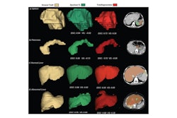
The traditional protocol included the following steps: Upper lung border at the first rib's upper edge; lower border below diaphragm (technician-dependent), covering apex to base. The precise protocol meanwhile included the following: Upper border aligns with the first rib (0 mm offset); lower border 35 mm (3 to 6 years) or 40 mm (7 to 14 years) below costophrenic angle.
The precise protocol led to z-axis coverage being reduced by 4.7% in children ages 3 to 6 years and 6.1% for children ages 7 to 14 years. It also yielded a less than 5% probability of missed lung parenchyma for both upper and lower boundaries using the optimized protocol.
The precise protocol also led to significant radiation dose reductions in both younger and older children.
Dose reductions in children after implementation of precise protocol | ||
Measure | Ages 3 to 6 years | Ages 7 to 14 years |
Effective dose | -19.2% | -17.0% |
Dose-length product | -17.7% | -17.0% |
The study authors also highlighted the protocol reducing internal and external variability among radiologic technologists.
“Overall, using this strategy, a satisfactory chest CT scan can be obtained, and the radiation dose can be minimized,” they wrote.
The authors also called for multi-center validation for their protocol and suggested that AI-driven automation “could further enhance clinical adoption.”
Read the full study here.