The RSNA has published an update to the CT Colonography Reporting and Data System (C-RADS) in Radiology, the first since the measure's implementation in 2005.
C-RADS is a classification scheme for CT colonography (CTC) findings, with C-RADS version 2023 including updates on the scheme used for colorectal and extracolonic findings on CTC, the RSNA said. Specifically, the update introduces a new subcategory, C2b, for mass-like diverticular strictures that are likely benign.
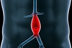
Graphical abstract of the C-RADS update. Image courtesy of RSNA.
Additionally, the update simplifies extracolonic classification by combining E1 and E2 categories into an updated extracolonic category of E1/E2, since no additional follow-up is required, irrespective of whether a finding is considered a normal variant (E1) or an otherwise clinically unimportant finding (E2), RSNA said.
“We hope this update encourages wider adoption of CTC and further standardizes the reporting and management of colonic and extracolonic findings using a simplified, clinically useful standardized lexicon and reporting structure for CTC,” wrote lead author Judy Yee, MD, of Albert Einstein College of Medicine in New York City, and colleagues.
The full article can be found here.


![Images show the pectoralis muscles of a healthy male individual who never smoked (age, 66 years; height, 178 cm; body mass index [BMI, calculated as weight in kilograms divided by height in meters squared], 28.4; number of cigarette pack-years, 0; forced expiratory volume in 1 second [FEV1], 97.6% predicted; FEV1: forced vital capacity [FVC] ratio, 0.71; pectoralis muscle area [PMA], 59.4 cm2; pectoralis muscle volume [PMV], 764 cm3) and a male individual with a smoking history and chronic obstructive pulmonary disorder (COPD) (age, 66 years; height, 178 cm; BMI, 27.5; number of cigarette pack-years, 43.2, FEV1, 48% predicted; FEV1:FVC, 0.56; PMA, 35 cm2; PMV, 480.8 cm3) from the Canadian Cohort Obstructive Lung Disease (i.e., CanCOLD) study. The CT image is shown in the axial plane. The PMV is automatically extracted using the developed deep learning model and overlayed onto the lungs for visual clarity.](https://img.auntminnie.com/mindful/smg/workspaces/default/uploads/2026/03/genkin.25LqljVF0y.jpg?auto=format%2Ccompress&crop=focalpoint&fit=crop&h=100&q=70&w=100)


![Images show the pectoralis muscles of a healthy male individual who never smoked (age, 66 years; height, 178 cm; body mass index [BMI, calculated as weight in kilograms divided by height in meters squared], 28.4; number of cigarette pack-years, 0; forced expiratory volume in 1 second [FEV1], 97.6% predicted; FEV1: forced vital capacity [FVC] ratio, 0.71; pectoralis muscle area [PMA], 59.4 cm2; pectoralis muscle volume [PMV], 764 cm3) and a male individual with a smoking history and chronic obstructive pulmonary disorder (COPD) (age, 66 years; height, 178 cm; BMI, 27.5; number of cigarette pack-years, 43.2, FEV1, 48% predicted; FEV1:FVC, 0.56; PMA, 35 cm2; PMV, 480.8 cm3) from the Canadian Cohort Obstructive Lung Disease (i.e., CanCOLD) study. The CT image is shown in the axial plane. The PMV is automatically extracted using the developed deep learning model and overlayed onto the lungs for visual clarity.](https://img.auntminnie.com/mindful/smg/workspaces/default/uploads/2026/03/genkin.25LqljVF0y.jpg?auto=format%2Ccompress&crop=focalpoint&fit=crop&h=112&q=70&w=112)


