
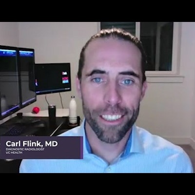
As a diagnostic radiologist at UC Health in Cincinnati, Carl Flink, MD, deals with many emergency cases. But on the night of January 2, he was part of a care case that caught the attention of many in the U.S.
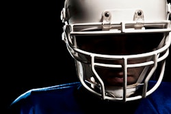
Flink was part of the emergency team that administered treatment to Buffalo Bills player Damar Hamlin, who suffered an on-field cardiac event after taking a hard hit. After receiving initial treatment on the football field, Hamlin was rushed to UC Health, where he made a full recovery.
Flink said while it is "wonderful" for the team to receive recognition from the NFL for its heroic actions, emergency radiologists can learn a thing or two from this event and apply lessons to their practices and workflows.


![Images show the pectoralis muscles of a healthy male individual who never smoked (age, 66 years; height, 178 cm; body mass index [BMI, calculated as weight in kilograms divided by height in meters squared], 28.4; number of cigarette pack-years, 0; forced expiratory volume in 1 second [FEV1], 97.6% predicted; FEV1: forced vital capacity [FVC] ratio, 0.71; pectoralis muscle area [PMA], 59.4 cm2; pectoralis muscle volume [PMV], 764 cm3) and a male individual with a smoking history and chronic obstructive pulmonary disorder (COPD) (age, 66 years; height, 178 cm; BMI, 27.5; number of cigarette pack-years, 43.2, FEV1, 48% predicted; FEV1:FVC, 0.56; PMA, 35 cm2; PMV, 480.8 cm3) from the Canadian Cohort Obstructive Lung Disease (i.e., CanCOLD) study. The CT image is shown in the axial plane. The PMV is automatically extracted using the developed deep learning model and overlayed onto the lungs for visual clarity.](https://img.auntminnie.com/mindful/smg/workspaces/default/uploads/2026/03/genkin.25LqljVF0y.jpg?auto=format%2Ccompress&crop=focalpoint&fit=crop&h=112&q=70&w=112)


