Photon-counting CT (PCCT) helps clinicians evaluate lung structure and function better than standard CT, according to a study published July 11 in Radiology.
The study results are exciting for improving patient care by reducing radiation exposure and for getting more out of CT imaging, noted a research group led by Sarah Scharm, MD, of Hannover Medical School in Germany.
"[Our] proposed photon-counting CT protocol is promising for a dose-efficient, robust, simultaneous acquisition and comprehensive evaluation of lung morphologic structure and function within a single pair of scans," the team wrote.
Chest CT is the gold standard for the evaluation of lung disease and for tracking disease progression, the researchers noted. But CT exams of lung function and blood flow require particular protocols that can't be combined. That's where PCCT comes in, offering high image quality, lower radiation dose compared to standard CT, better resolution, and spectral imaging options.
"The improvement in the contrast-to-noise ratio and spatial resolution of the pulmonary blood volume images was substantial," said corresponding author Hoen-oh Shin, MD, also of Hannover Medical School, in a statement released by the RSNA. "In my opinion, the most important advantage is the significantly improved spectral resolution, which enables new applications such as functional imaging of the lungs with CT."
Scharm and colleagues developed a chest imaging protocol that produces structural and functional data from CT images with PCCT. The protocol requires advanced software but no additional equipment.
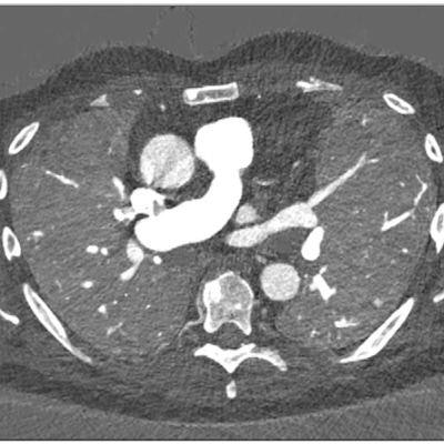
 Images in a 67-year-old male patient with slowly progressing idiopathic pulmonary fibrosis. The axial images show differences in the perfusion images generated with (A) photon-counting CT (PCCT) versus (B) standard dual-energy CT. The dose was slightly lower with PCCT (inspiration and expiration, respectively: CT dose index, 3.26 and 3.27 mGy; dose-length product, 126 and 128 mGy∙cm) versus dual-energy CT (inspiration and expiration, respectively: CT dose index, 4.52 and 3.28 mGy; dose-length product, 167.6 and 121.5 mGy∙cm). The technical advancement of PCCT results in improved spatial and contrast resolution. The faster spectral imaging of PCCT also allows for CT pulmonary angiography in addition to a stable, uniform parenchymal contrast. Furthermore, the morphologic images can be reconstructed with high spatial resolution (1024×1024). Images and caption courtesy of the RSNA.
The group conducted a study that included 166 patients with interstitial lung disease and post-COVID conditions; each individual underwent a contrast-enhanced inspiratory PCCT followed by expiratory PCCT after a delay of 5 minutes between November 2021 and June 2022.
The protocol simultaneously evaluated patients' lung structure, ventilation, vasculature, and perfusion of the parenchyma, demonstrating advantages over standard CT, the team reported.
It found the following:
- Mean dose-length product was 110.32 mGy ∙ cm* for the inspiration PCCT and 109.47 mGy ∙ cm, for the expiration PCCT.
- Mean CT dose index for the inspiration exam was 3.22 mGy and for the expiration exam was 3.09 mGy (both of which are less than the mean total radiation dose of 8 mGy to 12 mGy, the diagnostic reference standard).
The PCCT protocol shows promise for other lung imaging applications, such as preoperative evaluation of emphysema and perfusion defects in patients with chronic thromboembolic pulmonary hypertension, according to Scharm and colleagues. After surgery, the PCCT protocol could help clinicians evaluate the success of lung or stem cell transplant procedures.
The investigators plan to continue to refine the protocol so as to improve processing time, according to Shin.
"Regional ventilation and perfusion depend on patient position and gravity, among other factors," Shin said. "Further studies are needed to assess the dependence on position and depth of breathing, as well as the reproducibility of the measurements."