
Samsung Electronics subsidiary NeuroLogica has received clearance from the U.S. Food and Drug Administration (FDA) for a version of the company's OmniTom Elite mobile CT scanner with photon-counting detector (PCD) technology.
Called OmniTom Elite with PCD, the new system leverages the benefits of photon-counting CT on a mobile system with single-source CT technology and a single detector. The scanner can generate spectral CT images at multiple energy levels, according to the company.
Photon-counting CT scanners measure each individual x-ray photon as it passes through the patient's body. This differs from existing CT instrumentation, in which the scanner's detectors measure the total energy in many x-rays at once.
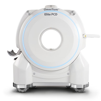
NeuroLogica's OmniTom Elite CT scanner has received 510(k) clearance for the addition of photon-counting detector (PCD) technology. Image courtesy of NeuroLogica.The Neurologica scanner's single x-ray source, when paired with a photon-counting CT detector, enables the generation of multiple sets of CT data acquired at the same time, using energy thresholds that can be configured without cross talk between images, according to the company. Photon-counting CT can lead to potentially more accurate visualization and segmentation of bone, as well as improved visualization of blood clots, plaque, hemorrhage, and intracranial tumors.
Photon-counting CT also has the potential to result in lower radiation dose and less use of contrast material.


![Images show the pectoralis muscles of a healthy male individual who never smoked (age, 66 years; height, 178 cm; body mass index [BMI, calculated as weight in kilograms divided by height in meters squared], 28.4; number of cigarette pack-years, 0; forced expiratory volume in 1 second [FEV1], 97.6% predicted; FEV1: forced vital capacity [FVC] ratio, 0.71; pectoralis muscle area [PMA], 59.4 cm2; pectoralis muscle volume [PMV], 764 cm3) and a male individual with a smoking history and chronic obstructive pulmonary disorder (COPD) (age, 66 years; height, 178 cm; BMI, 27.5; number of cigarette pack-years, 43.2, FEV1, 48% predicted; FEV1:FVC, 0.56; PMA, 35 cm2; PMV, 480.8 cm3) from the Canadian Cohort Obstructive Lung Disease (i.e., CanCOLD) study. The CT image is shown in the axial plane. The PMV is automatically extracted using the developed deep learning model and overlayed onto the lungs for visual clarity.](https://img.auntminnie.com/mindful/smg/workspaces/default/uploads/2026/03/genkin.25LqljVF0y.jpg?auto=format%2Ccompress&crop=focalpoint&fit=crop&h=100&q=70&w=100)


![Images show the pectoralis muscles of a healthy male individual who never smoked (age, 66 years; height, 178 cm; body mass index [BMI, calculated as weight in kilograms divided by height in meters squared], 28.4; number of cigarette pack-years, 0; forced expiratory volume in 1 second [FEV1], 97.6% predicted; FEV1: forced vital capacity [FVC] ratio, 0.71; pectoralis muscle area [PMA], 59.4 cm2; pectoralis muscle volume [PMV], 764 cm3) and a male individual with a smoking history and chronic obstructive pulmonary disorder (COPD) (age, 66 years; height, 178 cm; BMI, 27.5; number of cigarette pack-years, 43.2, FEV1, 48% predicted; FEV1:FVC, 0.56; PMA, 35 cm2; PMV, 480.8 cm3) from the Canadian Cohort Obstructive Lung Disease (i.e., CanCOLD) study. The CT image is shown in the axial plane. The PMV is automatically extracted using the developed deep learning model and overlayed onto the lungs for visual clarity.](https://img.auntminnie.com/mindful/smg/workspaces/default/uploads/2026/03/genkin.25LqljVF0y.jpg?auto=format%2Ccompress&crop=focalpoint&fit=crop&h=112&q=70&w=112)


