An incarcerated woman who underwent CT staging for breast cancer was found to have COVID-19 pneumonia, according to a case study to be published in the September issue of Radiology Case Reports.
The case yet again demonstrates how people infected with the SARS-CoV-2 virus can remain asymptomatic -- and how particular populations such as those in the prison system may be vulnerable, wrote a group led by Kevin Yoo, PhD, of Temple University Health System in Philadelphia.
"Infection with the novel severe acute respiratory syndrome coronavirus 2 ... may remain asymptomatic in the initial phase, leading to under-recognition and incidental detection on procedures for standard clinical indications," Yoo and colleagues noted.
It's common for patients with COVID-19 pneumonia to present with a now-recognized constellation of symptoms, such as fever, cough, and labored breathing, the group wrote. But there are also many patients who demonstrate no symptoms at all.
"However, there are others who may initially present as asymptomatic and remain as such," the group wrote. "Indeed, tracing asymptomatic and presymptomatic cases is vital in understanding the transmission of the virus, as well as curtailing further spread."
The case study described a 59-year-old incarcerated woman diagnosed with invasive ductal carcinoma in situ (DCIS) in 2016 who presented for follow-up after a 2019 mammogram showed that her cancer had progressed. The woman did not have fever, her oxygen saturation was normal, and she reported no chest pain or trouble breathing.
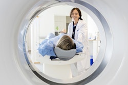
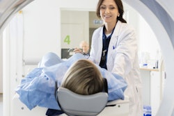
She underwent chest CT and her physicians identified incidental viral pneumonia findings of diffuse bilateral ground-glass opacities and a reverse halo sign in the right lower lobe. She then tested positive for COVID-19 on reverse transcription polymerase chain reaction (RT-PCR) assay.
The CT room was deep cleaned after her exam, the authors noted.
The case demonstrates yet again the need for healthcare professionals to be on the alert for COVID-19, no matter why a patient is presenting for imaging. The team outlined three takeaways from the case:
- Hospitals must prepare to encounter asymptomatic COVID-19 patients.
- Preparations should include providing staff with adequate personal protective equipment and perhaps reconfiguring the radiology department to safely house patients with suspected or confirmed COVID-19.
- Radiology departments should consider the financial and clinical implications of incidental COVID-19, such as increased risk of infection among staff and the need to delay imaging exams because of stricter infection control procedures.
"[Our] findings have important implications for infection control," Yoo and colleagues concluded. "There will inevitably be incidental detections in asymptomatic patients undergoing scans for other indications. ... Radiology departments and staff may face significant risk when performing diagnostic imaging on patients who may not be suspected of harboring COVID-19 due to the subclinical nature of incidental ... infections."