Autopsy misses injuries that can only be seen on postmortem CT for 17% to 21% of all injuries in those who died from firearms, pediatric trauma, or blunt force, according to a recent article on the National Institute of Justice website, NIJ.gov.
Researchers Sarah Lathrop, PhD, and Dr. Kurt Nolte worked with the New Mexico Office of the Medical Investigator to examine the potential of supporting or replacing forensic autopsies with postmortem CT.
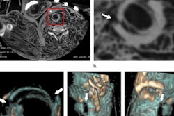
They evaluated 174 blunt force injury deaths, 205 firearm deaths, 65 pediatric trauma deaths, and 460 drug poisoning deaths that occurred between June 2011 and December 2013. In each case, a full autopsy and complete postmortem CT were performed.
There was strong agreement between autopsy and postmortem CT in assigning the cause of death, they found. In 85% of blunt force injury deaths, 99.5% of firearm fatalities, and 81.4% of pediatric trauma deaths, the cause of death determined by CT was correct, matching the autopsy. Agreement on cause of death was significantly less in drug poisoning deaths, ranging from 34.2% to 77.9%, with significantly less agreement in the deaths of people older than 40.
"In an ideal world, all medical examiners would have access to not only a CT scanner, but an experienced radiologist to interpret the results from the scans for them," Lathrop and Nolte wrote in their grant report. "In the majority of cases included in this study, postmortem CT, when paired with a thorough external examination, could supplant autopsy and would be of particular value in cases of family, religious, or cultural objections," they added.