The U.S. National Cancer Institute (NCI) has awarded a $2.9 million research grant to the University of California, Davis (UCD) for the development of CT to detect breast cancer.
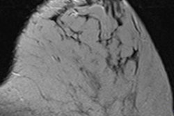
The research will be led by John Boone, PhD, a medical physicist and professor of radiology at UCD. Because breast cancer can present as both soft-tissue masses and microcalcifications, the new scanner needs to be able to detect both better than digital mammography, he said in a statement from the university.
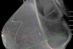
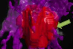
The study will compare mammography with breast CT to determine which modality is better at detecting breast lesions that are ultimately proved to be cancerous. The researchers will also compare contrast-enhanced breast CT with contrast-enhanced MRI. The study will include 400 women who have been found through standard-of-practice methods to have suspicious lesions that require breast biopsy.
If contrast-enhanced breast CT is equivalent to contrast-enhanced breast MRI, then CT would be a viable and more cost-effective option, Boone said. It would likely reduce the rate of negative biopsies and increase the positive predictive value of breast imaging overall, he added.
More than 300 patients have already received breast CT at UCD. The study team is also collaborating with the University of Pittsburgh, where a breast CT scanner designed by UCD has been used to scan an additional 300 patients.
The long-term goal of the research is to show that breast CT can improve detection rates in the screening population over those of mammography and tomosynthesis, he said.


![Axial images from unenhanced calcium score cardiac CT (left) and curved planar reformation images from CT angiography (right) show that higher long-term exposure to air pollution is associated with greater coronary artery calcium and more obstructive coronary artery disease (CAD). Top row: Images in a 68-year-old male patient with higher 10-year mean ambient air pollution exposure (7.9 μg/m3 for particulate matter measuring ≤2.5 μm in diameter [PM2.5] and 17.4 parts per billion [ppb] for NO2) with extensive CAD (coronary artery calcium score [CACS] >1,000 and obstructive CAD [≥70% diameter stenosis]). Bottom row: Images in a 57-year-old female patient with lower 10-year mean ambient air pollution exposure (6.3 μg/m3 for PM2.5 and 4.6 ppb for NO2) with no CAD (CACS = 0 and no obstructive stenosis).](https://img.auntminnie.com/mindful/smg/workspaces/default/uploads/2026/06/hanneman.r6SMLzkezo.png?auto=format%2Ccompress&fit=crop&h=100&q=70&w=100)


![Axial images from unenhanced calcium score cardiac CT (left) and curved planar reformation images from CT angiography (right) show that higher long-term exposure to air pollution is associated with greater coronary artery calcium and more obstructive coronary artery disease (CAD). Top row: Images in a 68-year-old male patient with higher 10-year mean ambient air pollution exposure (7.9 μg/m3 for particulate matter measuring ≤2.5 μm in diameter [PM2.5] and 17.4 parts per billion [ppb] for NO2) with extensive CAD (coronary artery calcium score [CACS] >1,000 and obstructive CAD [≥70% diameter stenosis]). Bottom row: Images in a 57-year-old female patient with lower 10-year mean ambient air pollution exposure (6.3 μg/m3 for PM2.5 and 4.6 ppb for NO2) with no CAD (CACS = 0 and no obstructive stenosis).](https://img.auntminnie.com/mindful/smg/workspaces/default/uploads/2026/06/hanneman.r6SMLzkezo.png?auto=format%2Ccompress&fit=crop&h=112&q=70&w=112)


