Dear CT Insider,
Evaluating for in-stent restenosis with CT has got to be one of radiology's toughest jobs, given the blinding white attenuation caused by many metallic stents on CT scans. But radiologists from Japan are hoping to boost their odds of evaluating stents with the aid of digital subtraction CT angiography (CTA), using a technique that works even for the smallest stents not currently recommended for coronary CTA.
In-stent restenosis isn't all that common, but it's not a job that can be skipped over either. Find out what the new technique produced in this issue's Insider Exclusive, brought to you as an Insider subscriber before our other AuntMinnie.com readers can access it.
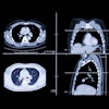
In the chest, it turns out that radiography has an important new job. OK, it's an old job: alerting physicians of the need for chest CT. In a study that looked at tens of thousands of radiographs in which radiologists saw something amiss, follow-up CT revealed serious abnormalities in a surprising percentage of referred cases, including plenty of previously unsuspected lung cancers.
The findings are not only surprising, they serve as a potent reminder of the limits of cost control at a time when requests for additional imaging are being heavily scrutinized. How many referrals are too many? And how many are too few, given the serious disease these CT scans are revealing? Food for thought, served up fresh here.
Lung cancer can spread via the airways, not just the blood, concludes new research from the University of Ottawa, which looked at the phenomenon of aerogenous spread of primary tumors within the lungs. Not only did these researchers find evidence of metastasis through this pathway, they offer great tips for finding patterns on CT that show it has occurred. Find out more here.
In a bit of good news on the oncology front, the American Cancer Society recently announced that cancer deaths have dropped by 22% over the past two decades. Death rates from breast cancer are down 35% from peak rates, while death rates from prostate and colorectal cancer are down by 47%. Learn more here.
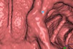
In CT colonography (also known as virtual colonoscopy), the use of dual-energy monochromatic datasets is improving the ability of computer-aided detection algorithms to spot lesions and distinguish them from other materials. Get the whole story from researchers at Massachusetts General Hospital by clicking here.
Moving on to the appendix, researchers from Turkey reported at RSNA 2014 on their use of a compression CT technique that doubled the number of diagnoses of acute appendicitis compared to ultrasound. Read more in this report.
In coronary artery news, a definitive new study from Korea found that dual-energy coronary CT angiography and CT myocardial perfusion imaging together improved both the identification and discrimination of ischemia-causing coronary artery stenoses over CTA alone. Get the whole story here.
And if you're wondering how often to follow up those nonsolid lesions in chest CT with additional imaging, you can relax a little. According to a report from Dr. Claudia Henschke, PhD, and her group's massive lung cancer database, these lesions actually harbor few cancers, and it's OK to wait a year rather than three months to follow up, as you'll learn by clicking here.
Finally, we invite you to scroll down through the links below for all of the news relating to radiology's most powerful and versatile modality, right here in your CT Community.

