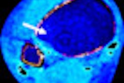
Image enhancement developer ContextVision is collaborating with Dr. Lars Borgen and Buskerud University College in Norway to explore technologies to enhance images in cases of reduced radiation dose.
Borgen used ContextVision's 2D and 3D filters to compare the effects of reduced dose in abdominal CT images. According to the company, the study's results show that ContextVision's 3D filter improves image quality compared to unfiltered and 2D filtered images.
For patients with a body mass index of less than 30 kg/m2, the 3D filter enabled 50% dose reduction in images, with comparable quality to full-dose images. Researchers rated all image quality criteria as superior for 3D filtered images compared to reduced-dose, baseline, and 2D filtered images.
Borgen said the study supports the use of highly sophisticated 3D image processing algorithms to reduce radiation from CT exams by as much as 50%.


![Images show the pectoralis muscles of a healthy male individual who never smoked (age, 66 years; height, 178 cm; body mass index [BMI, calculated as weight in kilograms divided by height in meters squared], 28.4; number of cigarette pack-years, 0; forced expiratory volume in 1 second [FEV1], 97.6% predicted; FEV1: forced vital capacity [FVC] ratio, 0.71; pectoralis muscle area [PMA], 59.4 cm2; pectoralis muscle volume [PMV], 764 cm3) and a male individual with a smoking history and chronic obstructive pulmonary disorder (COPD) (age, 66 years; height, 178 cm; BMI, 27.5; number of cigarette pack-years, 43.2, FEV1, 48% predicted; FEV1:FVC, 0.56; PMA, 35 cm2; PMV, 480.8 cm3) from the Canadian Cohort Obstructive Lung Disease (i.e., CanCOLD) study. The CT image is shown in the axial plane. The PMV is automatically extracted using the developed deep learning model and overlayed onto the lungs for visual clarity.](https://img.auntminnie.com/mindful/smg/workspaces/default/uploads/2026/03/genkin.25LqljVF0y.jpg?auto=format%2Ccompress&crop=focalpoint&fit=crop&h=112&q=70&w=112)