(Booth 2815) Medrad of Warrendale, PA, will highlight new software capabilities for its Stellant CT contrast injection system.
P3T Cardiac P3T Cardiac computes individual contrast injection protocols and scan timing for cardiac CT exams. Medrad has submitted an additional 510(k) submission for P3T Cardiac to the U.S. Food and Drug Administration for use in CT pulmonary angiography when used on studies to rule out pulmonary embolism, and clearance is pending.
The company has received 510(k) clearance for P3T Abdomen, which automates the calculation of protocols for CT abdominal (i.e. liver, kidney, and pancreas) imaging.
The Medrad Informatics Solution is a new product that automatically records injection data; the data are then sent to a Web site, label printer, and finally to the RIS. It is scheduled for release in Q1 2009. In future versions of the product, radiologists will be able to view a DICOM image of their injection protocol data in their PACS along with their patient’s image set. RIS integration is also planned in future phases.
In addition, Medrad will feature its XDS Extravasation Detector safety accessory that uses radiofrequency wave technology to detect mild extravasations. A contrast injection is automatically halted before moderate to severe damage can occur. This accessory has received FDA 510(k) clearance and has been available since July 2007.


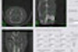


![Axial images from unenhanced calcium score cardiac CT (left) and curved planar reformation images from CT angiography (right) show that higher long-term exposure to air pollution is associated with greater coronary artery calcium and more obstructive coronary artery disease (CAD). Top row: Images in a 68-year-old male patient with higher 10-year mean ambient air pollution exposure (7.9 μg/m3 for particulate matter measuring ≤2.5 μm in diameter [PM2.5] and 17.4 parts per billion [ppb] for NO2) with extensive CAD (coronary artery calcium score [CACS] >1,000 and obstructive CAD [≥70% diameter stenosis]). Bottom row: Images in a 57-year-old female patient with lower 10-year mean ambient air pollution exposure (6.3 μg/m3 for PM2.5 and 4.6 ppb for NO2) with no CAD (CACS = 0 and no obstructive stenosis).](https://img.auntminnie.com/mindful/smg/workspaces/default/uploads/2026/06/hanneman.r6SMLzkezo.png?auto=format%2Ccompress&dpr=2&fit=crop&h=167&q=70&w=250)


