Dear CT Insider,
Lung nodules that grow over time present a higher risk of malignancy than stable lesions, of course. But deciding what is significant growth in which lesions, when to biopsy, and when to stick to CT surveillance is a tricky business indeed. Nodule size, morphology, location, volume doubling time and other factors all play an important role in the decision. The best available measuring tool, automated volumetric measurement, can output significant size variability between scans that is not related to lesion growth.
In this issue's CT Insider Exclusive, we explore the ins and outs of nodule volumetry, informed by the latest studies. Along the way you'll find advice from a real-life CT Insider, Dr. Mathias Prokop from University Medical Center Utrecht in the Netherlands, who offers tips on how to assess significant growth while avoiding the pitfalls of automated volumetry.
Also on the topic of lung cancer, find out how researchers are using advances in intensity-modulated radiation therapy to zero in on tumor and spare surrounding healthy tissues better than ever. Learn about the frequency of bronchoalveolar cell carcinoma in CT-detected ground-glass opacities. However, PET/CT was found to be of limited utility in lymph node staging of lung cancer.
In late-breaking news, researchers from the Netherlands report on their comparison of 64-slice CT with conventional angiography for asessing in-stent restenosis. Learn about their results by clicking here.
Finally, a report by staff writer Shalmali Pal takes a look at three studies presented at this week's American Society for Therapeutic Radiology and Oncology (ASTRO) meeting in Los Angeles that examine the role CT plays in targeting and planning radiotherapy for breast, brain, and lung cancer.
You're invited to scroll through the stories below for the rest of the news in CT imaging.


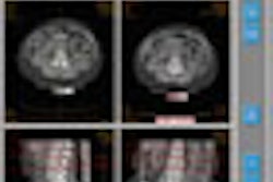


![Axial images from unenhanced calcium score cardiac CT (left) and curved planar reformation images from CT angiography (right) show that higher long-term exposure to air pollution is associated with greater coronary artery calcium and more obstructive coronary artery disease (CAD). Top row: Images in a 68-year-old male patient with higher 10-year mean ambient air pollution exposure (7.9 μg/m3 for particulate matter measuring ≤2.5 μm in diameter [PM2.5] and 17.4 parts per billion [ppb] for NO2) with extensive CAD (coronary artery calcium score [CACS] >1,000 and obstructive CAD [≥70% diameter stenosis]). Bottom row: Images in a 57-year-old female patient with lower 10-year mean ambient air pollution exposure (6.3 μg/m3 for PM2.5 and 4.6 ppb for NO2) with no CAD (CACS = 0 and no obstructive stenosis).](https://img.auntminnie.com/mindful/smg/workspaces/default/uploads/2026/06/hanneman.r6SMLzkezo.png?auto=format%2Ccompress&dpr=2&fit=crop&h=167&q=70&w=250)


