BARCELONA - In the early stage of acute coronary syndrome, 16-slice multidetector-row CT can locate the culprit lesion, according to a presentation this week at the European Society of Cardiology's World Congress of Cardiology.
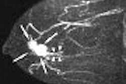
Researchers from University Hospital in Bordeaux, France compared 16-slice CT to 40-MHz catheter-based intravascular ultrasound (IVUS) in 20 consecutive patients who presented with acute coronary syndrome. Imaging studies were performed less than 48 hours after onset of symptoms.
Coronary plaque density was compared with echogenity assessed during three-vessel examination. Minimum lumen area and cross-sectional vessel area were manually traced for all significant lesions using multiplanar reconstructions that were oriented perpendicular to the longitudinal axis of the artery.
According to the results, 109 coronary segments were analyzed and 84 plaques were visualized by both techniques with good image quality. Multislice CT detected 19 of the 20 culprit lesions. Ten of those plaques were viewed in proximal segments of arteries on IVUS. Eight were hypoechogenic, five were hyperechogenic, and seven lesions were mixed.
Lead author Dr. Xavier Iriart said multislice CT proved effective in assessing lesions and their composition in carefully selected patients with stable coronary disease. However, plaque ruptures in five lesions, including ruptures in remote arteries in four patients, were not seen on CT, Iriart said.
Overall, measurements performed with multislice CT correlated well with IVUS (p < 0.001) although CT tended to overestimate vessel dimensions, he stated.
"While we can detect the lesion responsible for the symptoms, the clarity of the image is not yet ready for routine use in diagnosis," Iriart's group wrote in their electronic poster presentation. In comparison to IVUS, CT cannot ascertain the composition of the plaque as well as IVUS, especially for defining lesions that are likely to rupture, they explained.
Additionally, "the spatial resolution of the 16-slice CT remains a major drawback to quantitatively assess the severity of lesions in these unstable patients," the group stated.
By Edward SusmanAuntMinnie.com contributing writer
September 6, 2006
Related Reading
Dual-source imaging promises better CT scanning, June 15, 2006
Multidetector-row CT noninvasively measures coronary artery diameters, June 28, 2006
Copyright © 2006 AuntMinnie.com


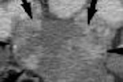


![Axial images from unenhanced calcium score cardiac CT (left) and curved planar reformation images from CT angiography (right) show that higher long-term exposure to air pollution is associated with greater coronary artery calcium and more obstructive coronary artery disease (CAD). Top row: Images in a 68-year-old male patient with higher 10-year mean ambient air pollution exposure (7.9 μg/m3 for particulate matter measuring ≤2.5 μm in diameter [PM2.5] and 17.4 parts per billion [ppb] for NO2) with extensive CAD (coronary artery calcium score [CACS] >1,000 and obstructive CAD [≥70% diameter stenosis]). Bottom row: Images in a 57-year-old female patient with lower 10-year mean ambient air pollution exposure (6.3 μg/m3 for PM2.5 and 4.6 ppb for NO2) with no CAD (CACS = 0 and no obstructive stenosis).](https://img.auntminnie.com/mindful/smg/workspaces/default/uploads/2026/06/hanneman.r6SMLzkezo.png?auto=format%2Ccompress&dpr=2&fit=crop&h=167&q=70&w=250)
