Does teleradiology increase the risk of malpractice liability for radiologists? It seems so, according to a study covered in our most highly viewed article last week.
After researchers from Harvard Medical School retrospectively reviewed medical malpractice claims over a 12-year period, they found that cases involving teleradiology tended to be more severe and result in higher indemnity payments than other radiology malpractice claims.
Evidence continues to build that AI can help reduce the number of interval breast cancers. Researchers from Turkey recently reported success after AI was implemented in Turkey’s national breast cancer screening program. Click here to learn more.
Our series on the rise of theranostics also returned this week. In part 2, learn what you need to know before venturing into theranostics for prostate cancer.
CT angiography (CTA) may be overused in the emergency department, according to another popular story. Although CTA has been increasingly used for patients with headache or dizziness, the rate of positive findings in these exams has decreased, according to the group from Massachusetts General Hospital. Click here for our report.
AI analysis of SPECT/CT myocardial perfusion imaging studies can predict if a patient will be hospitalized for heart failure, potentially enabling earlier interventions in these patients. Additionally, ChatGPT shows promise for referring patients to interventional radiology for treatment.
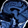
And a team from France has released beautiful brain MRI images from their 11.7-tesla scanner.
See the full list below of our most popular stories of the week:
- Does teleradiology increase malpractice risk for radiologists?
- AI shows promise in detecting interval breast cancers
- The rise of theranostics: Part 2 -- Moving into communities
- Is CTA overused in emergency department?
- SPECT/CT algorithm predicts heart failure hospitalizations
- Can ChatGPT accurately refer patients to IR for treatment?
- French team unveils 11.7-tesla MRI scans of human brain
- ChatGPT performs well on radiation oncology patient care questions
- MRI shows preeclampsia’s effect on maternal heart, fetal brain
- Clinical value of some knee and hip MRIs questioned
- AIUM president-elect previews UltraCon 2024
- Abortion care limitations raise medicolegal concerns in IR
- Can AI help chest DDR achieve clinical adoption?
- 9 ways to ease the radiologist workforce shortage