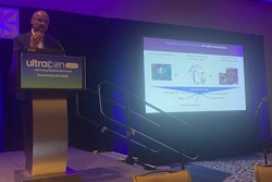
Attendees at this year’s UltraCon in Austin, TX, can look forward to the new research and technology to be highlighted -- as well as the solar eclipse that will take place during the conference.
UltraCon, formerly the American Institute of Ultrasound in Medicine (AIUM) annual meeting, from April 6 to 10 will feature research covering several areas in ultrasound, including point-of-care ultrasound (POCUS), elastography, contrast enhancement, and AI, among others.
“There’s that synergy that occurs when you get all these people together,” said David Jones, MD, president-elect of AIUM. “I think everybody is going to have a good experience, get a great education, and come out of it stimulated to know how they can use technology that very day and down the road, into the future.”
UltraCon will present opportunities for professional networking, educational enhancement, and reviewing technologies that could improve clinical practice. The conference will have events such as “Sono-Jeopardy,” industry presentations, and a “Shark Tank” event for competitors to pitch their ideas to an expert panel.
Also, AIUM will give attendees specialized glasses to view the eclipse on April 8. The eclipse will be able to be viewed in Austin around 1:30 p.m. local time. Jones, a self-described amateur astronomer, said when looking at eclipse dates about two years ago, he realized the 2024 eclipse would fall on a date when UltraCon would be held, as well as within the viewing range.
“Fortunately, we had these hotel dates set long before Hilton was paying attention,” Jones said. “If Mother Nature cooperates and the clouds are parted, people will be able to look safely at the eclipse."
And while AI continues to gain ground in imaging departments, Jones said the technology has a while to go before becoming a day-to-day part of clinical workflows. This includes making sure staff members have the necessary training in image interpretation and diagnosis when using AI.
“It’s making sure that people are adequately trained to use it properly,” he told AuntMinnie.com. “It’s also realizing that you still need to use your human reading skills to put things into context.”
Finally, Jones said that there is something for everybody when it comes to takeaways from the conference by subspecialty.
“If you’re a sonographer, you’re going to learn everything from how to image whatever part of the body is within your expertise to how to hold your body in a way that’s ergonomically safer,” he said. “If you’re a physician or a provider who interprets ultrasound images, you’re going to learn how to do that better.”