A team of researchers in Bangladesh has launched a dataset of x-ray images called FracAtlas that could be valuable for developing AI algorithms to diagnose bone fractures, according to a study published August 5 in Scientific Data.
The dataset includes 4,083 images that two expert radiologists and an orthopedist have manually annotated for bone fracture classification, localization, and segmentation, lead co-authors Iftekharul Abedeen and Md. Ashiqur Rahman of the Islamic University of Technology in Gazipur explained.
"We believe the dataset will be a valuable resource for researchers interested in developing and evaluating machine learning algorithms for bone fracture diagnosis," the group wrote.
Digital radiography is one of the most common and cost-effective methods for diagnosing bone fractures. The field is experiencing a boom in efforts to develop AI algorithms that can help overworked radiologists and underserved patients, yet most existing x-ray datasets are either small or lack proper annotation, according to the authors.
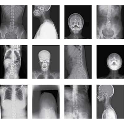
To address the knowledge gap, the group first collected 14,068 x-ray scans from three hospitals and diagnostic centers in Bangladesh taken between 2021 and 2022. They culled 4,083 normal and abnormal images of the hand, leg, hip, and shoulder regions, then identified the presence and number of fractures on each image.
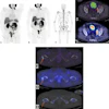
 Example of valid (left) vs outlier (right) x-ray images. All the scans were manually filtered based on the parts of the body present in the scan, clarity of the scans, and resolution. The scans containing only arm, shoulder, leg, and hip regions were accepted. Image and caption courtesy of Scientific Data through CC BY 4.0.
Next, Abedeen and colleagues manually annotated the images using makesense.ai, a free, open-source tool, marking regions of interest. After the annotation process, they validated the dataset for both fracture localization and segmentation using YOLO (You Only Look Once) object detection models.
In total, out of 4,083 images, the dataset contained 717 images that had 922 instances of fractures. The age of subjects in the dataset ranged from eight months to 78 years old.
Since most prominent AI algorithms for bone fracture classification, localization, and segmentation have used private datasets -- due to the unavailability of publicly accessible datasets -- FracAtlas fills a need, according to the authors, who noted that the FracAtlas dataset is available for free public download through Figshare.
The full article is available here.