A new radiation exposure shield designed to provide total-body protection could allow some radiologists to ditch burdensome lead aprons, according to a study published March 21 in the Journal of the Society for Cardiovascular Angiography and Interventions.
A team led by Dr. David Rizik of HonorHealth Heart Group in Scottsdale, AZ, tested the use of a shielding system called Protego (Image Diagnostics) during routine procedures at their hospital's cardiac catheterization lab. Compared with leaded apparel and drop-down shields, the new system provided superior protection, the group found.
"This shielding approach can eliminate the need for orthopedically burdensome personal leaded apparel and has the potential to reduce catheterization laboratory occupational health hazards," Rizik and colleagues wrote.
Studies have shown that interventional cardiologists and cardiac catheterization laboratory staff face significant occupational hazards related to ionizing radiation, with radiation exposure causing both direct tissue damage and risks related to DNA damage, namely cancer. Societies representing interventional radiologists have emphasized the need for workplace radiation protection, with the ultimate goal of achieving as close to zero exposure as possible, the authors wrote.
Early data has shown that the first iteration of the Protego shield provides high levels of protection. In this study, the investigators aimed to test its performance in a real-world clinical setting. They compared physician operator radiation exposure using the Protego shield in 25 cases versus standard protection (personal leaded apparel and a drop-down shield) in 25 cases during routine cardiac catheterization procedures. Exposures at both thyroid and waist levels were measured with a real-time dosimetry system and calculated on a mean-per-case basis.
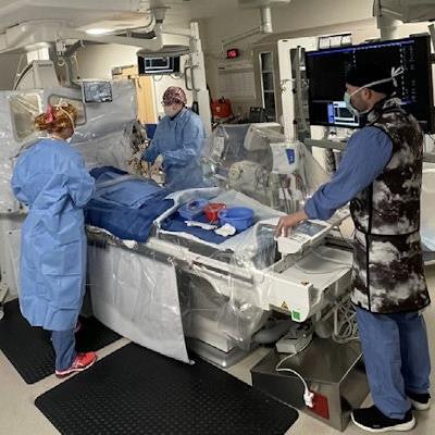
 The Protego shield in clinical operation. (A) Protego shield set up. (B) In clinical use. The shield casts a broad area of protection that may benefit all catheterization laboratory personnel. Image and caption courtesy of the Journal of the Society for Cardiovascular Angiography and Interventions through CC BY 4.0.
Radiation protection with the Protego was superior to standard methods, showing markedly lower radiation exposure at both the thyroid level (0.36 vs. 58.5 µSv) and the waist level (0.84 vs. 121.4 µSv). Zero total radiation exposure was documented in 68% of Protego cases, while standard protection did not achieve this coverage in a single case, the researchers reported.
"The magnitude of protection achieves state regulatory standards sufficient to allow operators to perform procedures without orthopedically burdensome lead aprons," Rizik and colleagues wrote.
In an accompanying editorial, Dr. Paul Fiorilli of the University of Pennsylvania in Philadelphia and Dr. Andrew Goldsweig of Baystate Medical Center in Springfield, MA, wrote that the minuscule level of operator radiation exposure with the Protego system is lower than levels ever reported previously with any radiation protection strategy. They did acknowledge that health systems may balk at incorporating novel protection systems such as Protego -- owing to the upfront costs of implementing it -- but they said that the safety of staff should trump those concerns.
"Physicians and physician-led organizations such as the Society for Cardiovascular Angiography and Interventions should demand this level of protection for themselves and their staff," they concluded.
Disclosure: Co-author Dr. James Goldstein declares interest in ECLS, Inc., a company that licenses technology to Image Diagnostics.