Investigators from Germany say that their prototype dark-field chest x-ray system may be able to help differentiate between emphysema and fibrosis, as discussed in a case published August 18 in Radiology: Cardiothoracic Imaging.
An 83-year-old man presented recently at their hospital with combined pulmonary fibrosis and emphysema (CPFE), wrote first authors Dr. Florian Gassert of the diagnostic and interventional radiology department at the Technical University of Munich, and physicist Theresa Urban.
A chest CT scan showed advanced upper lung mixed centrilobular and paraseptal emphysema and lower lung-predominant peripheral fibrotic changes with honeycombing, indicating an interstitial pneumonia pattern. In addition, pulmonary function tests showed combined obstructive to forced vital capacity and restrictive lung changes, with impaired lung diffusion capacity.
The researchers used the prototype system to acquire both a dark-field chest x-ray image and a conventional attenuation-based chest x-ray image of the patient in a single scan.
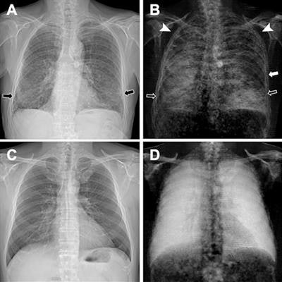
Figure 2: (A, C) Conventional attenuation-based chest x-rays and (B, D) dark-field chest x-rays in an 83-year-old man diagnosed with combined pulmonary fibrosis and emphysema (upper row) and a healthy 33-year-old man (lower row). Image courtesy of Radiology: Cardiothoracic Imaging.The patient (Fig 2: A, B) demonstrated substantially decreased dark-field signal in the upper lungs (left more than right), where emphysema is most severe, and inhomogeneous but decreased lower lung signal (due to presence of fibrosis) when compared with a healthy subject (Fig 2: C, D). The stronger manifestation of fibrosis in the right lower lobe compared with the left lower lobe, as was seen on CT images, corresponded well to the asymmetric reduction of dark-field signal in the lower fields with a right-sided predominance (Fig 2B), the authors wrote.
"As both fibrosis and emphysema lead to a reduction of dark-field signal -- due to a smaller number of intact alveoli -- the assignment of signal loss to one of the two pathologic conditions is hardly possible from the [dark-field chest x-ray] alone," the researchers wrote.
However, the combination of conventional and dark-field images might allow for the differentiation of emphysema and fibrosis, especially as the images are perfectly coregistered, they suggested.
While the dark-field signal appearance of fibrosis needs to be further investigated, the researchers are optimistic, given that first published studies have shown that dark-field chest x-ray allows for the qualitative and quantitative assessment of emphysema, Gassert and Urban concluded.

![Examples of ultrasound findings and techniques. (A) Images in a 39-year-old male patient with a mass in the left thigh. The mass is heterogeneous on the B-mode US image (compared with the patient in D) and showed increased microvascularity (superb microvascular imaging [SMI]) and shear-wave elastography (SWE) values. Undifferentiated pleomorphic sarcoma was diagnosed at biopsy (with pleomorphic rhabdomyosarcoma in surgical specimen). (B) Images in an 18-year-old male patient with a mass in the left leg. The mass is hypoechoic on the B-mode image, with no other findings suggestive of malignancy. The lesion is in contact with the cortex of the tibia, which is slightly irregular. CT revealed a doubtful anteromedial tibial erosion. The microvascular study demonstrated high vascularization, suggestive of malignancy. Periosteal Ewing sarcoma was diagnosed with both histologic and immunohistochemical confirmation. (C) Images in a 69-year-old female patient with a lump growing on the outside of the left leg. Multiple SWE examinations were performed (please note the high values obtained in the measurements, whereas the color map highlights the stiffness relative to adjacent tissues). SMI showed areas of increased vascularization to target for sampling. Undifferentiated spindle cell sarcoma was diagnosed at biopsy, with residual leiomyosarcoma in the surgical specimen after neoadjuvant therapy. (D) Images in a 56-year-old female patient with a mass in the right thigh. The mass is heterogeneous at both B-mode ultrasound (similar to patient A) and MRI (coronal T2-weighted spectral attenuated inversion recovery [SPAIR]; T1-weighted pre-contrast and postcontrast imaging), which even shows uptake after the administration of paramagnetic contrast material, which is traditionally suggestive of malignancy. Low values at SMI and elastography are suggestive of benignity. Spindle cell lipoma was diagnosed at biopsy, with atypical spindle cell lipomatous tumor in the surgical specimen.](https://img.auntminnie.com/mindful/smg/workspaces/default/uploads/2026/08/images-radiol250278fig2.APCFLSvX6p.jpg?auto=format%2Ccompress&fit=crop&h=100&q=70&w=100)


![Examples of ultrasound findings and techniques. (A) Images in a 39-year-old male patient with a mass in the left thigh. The mass is heterogeneous on the B-mode US image (compared with the patient in D) and showed increased microvascularity (superb microvascular imaging [SMI]) and shear-wave elastography (SWE) values. Undifferentiated pleomorphic sarcoma was diagnosed at biopsy (with pleomorphic rhabdomyosarcoma in surgical specimen). (B) Images in an 18-year-old male patient with a mass in the left leg. The mass is hypoechoic on the B-mode image, with no other findings suggestive of malignancy. The lesion is in contact with the cortex of the tibia, which is slightly irregular. CT revealed a doubtful anteromedial tibial erosion. The microvascular study demonstrated high vascularization, suggestive of malignancy. Periosteal Ewing sarcoma was diagnosed with both histologic and immunohistochemical confirmation. (C) Images in a 69-year-old female patient with a lump growing on the outside of the left leg. Multiple SWE examinations were performed (please note the high values obtained in the measurements, whereas the color map highlights the stiffness relative to adjacent tissues). SMI showed areas of increased vascularization to target for sampling. Undifferentiated spindle cell sarcoma was diagnosed at biopsy, with residual leiomyosarcoma in the surgical specimen after neoadjuvant therapy. (D) Images in a 56-year-old female patient with a mass in the right thigh. The mass is heterogeneous at both B-mode ultrasound (similar to patient A) and MRI (coronal T2-weighted spectral attenuated inversion recovery [SPAIR]; T1-weighted pre-contrast and postcontrast imaging), which even shows uptake after the administration of paramagnetic contrast material, which is traditionally suggestive of malignancy. Low values at SMI and elastography are suggestive of benignity. Spindle cell lipoma was diagnosed at biopsy, with atypical spindle cell lipomatous tumor in the surgical specimen.](https://img.auntminnie.com/mindful/smg/workspaces/default/uploads/2026/08/images-radiol250278fig2.APCFLSvX6p.jpg?auto=format%2Ccompress&fit=crop&h=112&q=70&w=112)