
"Scanxiety" among patients with serious disease is common, and it can be severe. But there are ways to soothe patient anxiety for imaging studies, according to research published online August 1 in Supportive Care in Cancer.
"Practical and psychosocial factors do affect the scan experience and are a possible target for interventions in future studies," wrote a team led by Dr. Kim Tam Bui of Concord Cancer Centre in Concord, Australia.
The term "scanxiety" was invented by a patient who wrote a 2011 Time Magazine article about the phenomenon. It's defined as "distress before, during, or after a cancer-related scan," and its incidence can range to up to 83%, the team wrote. Unfortunately, people with advanced cancer must undergo frequent imaging -- thus raising their risk of anxiety related to these exams.
"The experience of scanxiety may be exacerbated by profound implications of scan results on cancer treatment or expectations of prognosis," Bui and colleagues wrote.
The team conducted a study that included 222 advanced cancer patients who had undergone a CT scan within the past four months. Participants completed a survey between March 2019 and March 2020 through which they rated the presence and severity of their anxiety about imaging on a scale of 1 to 10.
The most common cancer types the researchers found were breast (37%), lung (19%), and colorectal (16%); most patients (82%) were less than one year post diagnosis. More than half (55%) of study participants experienced "scanxiety," and the mean anxiety severity score was 6 on a range of 1 to 10.
| Analysis of imaging exam anxiety prevalence and severity by patient factors | ||
| Factor | Prevalence | Severity (scale of 1 to 10) |
| Female vs. male | 65% vs. 38% | 6.2 vs. 5.4 |
| Inhabiting urban vs. rural setting | 50% vs. 66% | 6 vs. 5.7 |
| Breast cancer vs. other cancers | 68% vs. 46% | 6.1 vs. 5.7 |
| Cancer diagnosis less than a year before vs. more than a year before | 51% vs. 57% | 6.7 vs. 5.7 |
| Wait between scan and results of less than two days vs. more than two days | 43% vs. 59% | 6.7 vs. 5.7 |
| Clinical anxiety vs. no clinical anxiety | 89% vs. 50% | 7.2 vs. 5.6 |
| Clinical depression vs. no clinical depression | 73% vs. 54% | 5.9 vs. 6 |
What helped mitigate patients' scanxiety? The authors listed some of the top factors patients reported (scores of 7 or higher out of 10):
- Friendly staff
- Knowing what will happen during the scan
- Undergoing the CT scan close to home
- Having the scan at the hospital where one is undergoing treatment
- Being informed of wait time for the scan
- Getting results from one's own oncologist
This patient feedback suggests specific ways imaging providers could try to soothe anxiety about imaging, according to the authors.
"Research to improve the scan experience and reduce scanxiety ... could be guided by our study findings," they wrote. "Interventions to reduce scanxiety could ... incorporate strategies reported by our participants, including psychosocial support, or activities to distract, relax, or energize people around a scan."
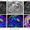

![Examples of ultrasound findings and techniques. (A) Images in a 39-year-old male patient with a mass in the left thigh. The mass is heterogeneous on the B-mode US image (compared with the patient in D) and showed increased microvascularity (superb microvascular imaging [SMI]) and shear-wave elastography (SWE) values. Undifferentiated pleomorphic sarcoma was diagnosed at biopsy (with pleomorphic rhabdomyosarcoma in surgical specimen). (B) Images in an 18-year-old male patient with a mass in the left leg. The mass is hypoechoic on the B-mode image, with no other findings suggestive of malignancy. The lesion is in contact with the cortex of the tibia, which is slightly irregular. CT revealed a doubtful anteromedial tibial erosion. The microvascular study demonstrated high vascularization, suggestive of malignancy. Periosteal Ewing sarcoma was diagnosed with both histologic and immunohistochemical confirmation. (C) Images in a 69-year-old female patient with a lump growing on the outside of the left leg. Multiple SWE examinations were performed (please note the high values obtained in the measurements, whereas the color map highlights the stiffness relative to adjacent tissues). SMI showed areas of increased vascularization to target for sampling. Undifferentiated spindle cell sarcoma was diagnosed at biopsy, with residual leiomyosarcoma in the surgical specimen after neoadjuvant therapy. (D) Images in a 56-year-old female patient with a mass in the right thigh. The mass is heterogeneous at both B-mode ultrasound (similar to patient A) and MRI (coronal T2-weighted spectral attenuated inversion recovery [SPAIR]; T1-weighted pre-contrast and postcontrast imaging), which even shows uptake after the administration of paramagnetic contrast material, which is traditionally suggestive of malignancy. Low values at SMI and elastography are suggestive of benignity. Spindle cell lipoma was diagnosed at biopsy, with atypical spindle cell lipomatous tumor in the surgical specimen.](https://img.auntminnie.com/mindful/smg/workspaces/default/uploads/2026/08/images-radiol250278fig2.APCFLSvX6p.jpg?auto=format%2Ccompress&fit=crop&h=100&q=70&w=100)


![Examples of ultrasound findings and techniques. (A) Images in a 39-year-old male patient with a mass in the left thigh. The mass is heterogeneous on the B-mode US image (compared with the patient in D) and showed increased microvascularity (superb microvascular imaging [SMI]) and shear-wave elastography (SWE) values. Undifferentiated pleomorphic sarcoma was diagnosed at biopsy (with pleomorphic rhabdomyosarcoma in surgical specimen). (B) Images in an 18-year-old male patient with a mass in the left leg. The mass is hypoechoic on the B-mode image, with no other findings suggestive of malignancy. The lesion is in contact with the cortex of the tibia, which is slightly irregular. CT revealed a doubtful anteromedial tibial erosion. The microvascular study demonstrated high vascularization, suggestive of malignancy. Periosteal Ewing sarcoma was diagnosed with both histologic and immunohistochemical confirmation. (C) Images in a 69-year-old female patient with a lump growing on the outside of the left leg. Multiple SWE examinations were performed (please note the high values obtained in the measurements, whereas the color map highlights the stiffness relative to adjacent tissues). SMI showed areas of increased vascularization to target for sampling. Undifferentiated spindle cell sarcoma was diagnosed at biopsy, with residual leiomyosarcoma in the surgical specimen after neoadjuvant therapy. (D) Images in a 56-year-old female patient with a mass in the right thigh. The mass is heterogeneous at both B-mode ultrasound (similar to patient A) and MRI (coronal T2-weighted spectral attenuated inversion recovery [SPAIR]; T1-weighted pre-contrast and postcontrast imaging), which even shows uptake after the administration of paramagnetic contrast material, which is traditionally suggestive of malignancy. Low values at SMI and elastography are suggestive of benignity. Spindle cell lipoma was diagnosed at biopsy, with atypical spindle cell lipomatous tumor in the surgical specimen.](https://img.auntminnie.com/mindful/smg/workspaces/default/uploads/2026/08/images-radiol250278fig2.APCFLSvX6p.jpg?auto=format%2Ccompress&fit=crop&h=112&q=70&w=112)