It can be difficult to adapt a technique that was developed in an academic venue for use in a community hospital environment. Technology, personnel, and budgetary considerations often limit the offerings that can be provided locally. Sometimes, however, the addition of an element as simple as a commercial software package can expand the range of clinical treatments that a community hospital can deliver.
A group from Aultman Hospital in Canton, OH, has taken a research-based SPECT/CT immunoscintigraphy-guided radiation therapy methodology developed at Case Western Reserve University School of Medicine in Cleveland, and successfully adopted the protocol into its practice. The team presented its findings on the methodology in a poster presentation at the Society of Nuclear Medicine (SNM) conference in Toronto last June.
The protocol was developed from a cohort of 239 patients at Case treated with brachytherapy utilizing SPECT images fused with CT to direct treatment planning for dose escalation to biologic tumor volume (BTV), according to the authors.
"Research-based SPECT/CT immunoscintigraphy-guided radiation treatment methodology was adopted by cross-functional teams from nuclear medicine and radiation oncology within a community hospital setting for assimilation into routine clinical use," they wrote.
For purposes of the study, a patient was referred to Aultman Hospital for radiation treatment following repeated negative biopsy and subsequent saturation biopsy. Images were acquired on a Philips Medical Systems (Andover, MA) ADAC SPECT system using indium-111 capromab pendetide (ProstaScint, manufactured by Cytogen of Princeton, NJ) and technetium-99m-labeled red blood cells as radioisotopes. A GE Healthcare (Chalfont St. Giles, U.K.) LightSpeed CT scanner was used for anatomical imaging.
SPECT images were acquired in a 128 x 128 matrix at 64 steps per head and 55 seconds per step from the top of the patient's head to the midthigh using ProstaScint (5-6 mCi) only on one day and both isotopes (ProstaScint scan followed by Tc-99m RBC, 4-5 mCi) on the second day of scanning. The SPECT and CT images were then fused using MIM (MIMvista, Cleveland), a medical image review application that includes image fusion capabilities for tomographic images from multiple modalities.
"The resulting SPECT/CT image was evaluated to guide radiation therapy dose escalation to areas of radioimmunoscintigraphy uptake, with concomitant dose sparing of normal tissues associated with increased treatment-related morbidity," the authors wrote.
 |
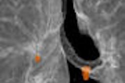
| Typical example of ProstaScint scans and dose escalation with brachytherapy. Image and caption courtesy of Dr. Rodney Ellis, Aultman Hospital, Canton, OH. |
The researchers correlated the areas of uptake on the SPECT/CT images with multicore biopsy, limited to five of eight cores on the right side of the prostate. They reported that the areas of uptake (focal activity in high anterior right base and extending to lateral right apex) were associated with positive findings from the saturation biopsy, and used the data to target dose escalation with brachytherapy.
"Both biopsies obtained at the time of the implant from the dominate lesion in the right anterior base were positive for adenocarcinoma with a Gleason score of 6 (3+3)," the authors wrote. "A single biopsy of the right lateral apex was negative, likely due to small volume, and heavy preoperative treatment with two months of hormonal therapy and five weeks of external beam radiation."
The authors noted that their application of ProstaScint is not contained in the prescribing information for the radiopharmaceutical. Also, they stated that the original studies for U.S. Food and Drug Administration approval of the agent did not evaluate the capability of ProstaScint to image disease within the prostate gland, or to direct the placement of brachytherapy sources or otherwise guide therapy to the prostate gland. Use of the agent does show promise for these applications, however.
"Functional imaging with ProstaScint may allow dose intensification to tumor areas expressing prostate-specific membrane antigen and allow dose reduction to normal areas," the authors wrote.
By Jonathan S. Batchelor
AuntMinnie.com staff writer
August 15, 2005
Related Reading
Pinhole SPECT shows promise for arthritis imaging, July 22, 2005
UMC Utrecht launches high-resolution SPECT, July 21, 2005
Can SPECT/CT revitalize nuclear medicine? July 7, 2005
UCLA launches brain SPECT venture, June 20, 2005
Abnormal SPECT predicts worse cardiovascular outcome in minorities, May 4, 2005
Copyright © 2005 AuntMinnie.com