FDG-PET offers an accurate way to assess cervical tumor volumes post-treatment, but the prognosis is particularly grim for patients who exhibit residual disease on PET scans after brachytherapy, according to radiation oncologists from the Mallinckrodt Institute of Radiology in St. Louis.
"This work is part of a larger project in which we set forth ... to use FDG-PET scans for brachytherapy treatment planning, instead of CT or MRI," explained Dr. Perry Grigsby in a presentation at the 2005 American Brachytherapy Society (ABS) meeting in San Francisco. "We didn't intend to look at specific changes in the volume of the tumor based on FDG-PET, but it became evident that there were significant changes in the volume, and that this has significant implications."
For their substudy, Grigsby's group enrolled 32 women with invasive cervical cancer (29 had squamous cell carcinoma). The median tumor volume for stage I cancers was 54 cc, for stage II it was 79 cc, and 176 cc for stage III. Patients were all treated with external-beam radiation therapy, brachytherapy, and chemotherapy.
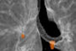
The group used an in-house software program that detected tumor boundaries on the PET scans. CT and MRI measurements of tumor volume were performed first and used to validate PET tumor volumes (see image below).
 |
"The boundary of the tumor was determined to be that voxel that had 40% of the peak FDG activity within the tumor volume. Then we could do a 3D volume calculation," Grigsby said.
FDG-PET volume measurements were first generated at the time of diagnosis. The patients underwent six fractions of brachytherapy during the course of their external-beam radiation treatment. PET scans were then done two, four, and six weeks into therapy with the brachytherapy implant applicator in place. Finally, follow-up PET scans were done three months after treatment.
According to the results, successful treatment took place when the tumor volume decreased to about 8% of its original size by the sixth week of therapy. Of the 32 patients, only four had any measurable residual volume, and all of those women died of their disease, Grigsby said.
 |
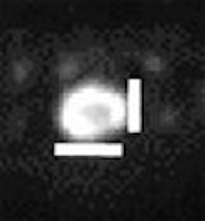
| Graphic illustrates survival versus residual volume. Images courtesy of Dr. Perry Grigsby. |
"Our conclusion from this study would be that PET can be used to determine tumor volume quite accurately," he said. "The tumor volume decreases exponentially during the course of therapy either with time or dose. Residual tumor volume at three months after completion of therapy is a very poor prognostic factor."
FDG uptake at three months can be taken as a sign of local resistance to radiation, rendering additional brachytherapy treatment fairly useless, he said. In those patients, Grigsby recommended hysterectomy.
The results of FDG-PET scans can also be used to recommend adjuvant surgery, Grigsby said in response to a question from Dr. D. Jeffrey Demanes, the ABS session moderator.
A session attendee asked Grigsby if FDG uptake because of inflammation was a problem in this study, as the latter can skew tumor boundaries.
"In the world of nuclear medicine, the issue of uptake of FDG in inflammation is often discussed," Grigsby responded. "In my experience, inflammatory issues in the patient with pelvic cancer almost never happen. Inflammation happens in colorectal patients, it happens in lung cancer, but it doesn't seem to happen in this group of patients."
Other advantages of PET in cervical cancer include the ability to determine the status of the lymph nodes at diagnosis, Grigsby told AuntMinnie.com. More importantly, this procedure is covered by Medicare, he said.
By Shalmali Pal
AuntMinnie.com staff writer
July 14, 2005
Related Reading
CMS provides details on new PET coverage, April 8, 2005
Few older cervical carcinoma patients get aggressive treatment, January 31, 2005
Copyright © 2005 AuntMinnie.com