Dual-tracer PET/CT appears valuable for improving tumor staging in patients with hepatocellular carcinoma being evaluated for treatment, according to a study published April 25 in JHEP Reports.
A group led by researchers at the University of Hong Kong in China found the approach detected 12% more disease in patients undergoing staging than CT or MRI and resulted in a change in treatment in a significant number of cases. The results validate the emerging technique, the group noted.
“In a large series of patients, we provided quantitative evidence for the use of combined F-18 FDG and C-11 acetate PET/CT in staging HCC patients,” wrote lead author Keith Wan Hang Chiu, MD, and colleagues.
Hepatocellular carcinoma (HCC) is the third most common cause of cancer-related deaths worldwide, accounting for over 800,000 deaths annually. Currently, CT and MRI are the imaging modalities of choice for assessing diagnosed patients, yet have been shown to miss small lesions or metastatic deposits, the authors explained.
Conversely, combined F-18 FDG and carbon-11 (C-11) acetate (dual-tracer) PET/CT has shown excellent sensitivity and specificity for detecting HCC in small studies, they added.
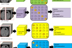
Thus, to provide more evidence on the approach, the researchers evaluated it for tumor staging and characterizing indeterminate lesions in 525 HCC patients. A majority (n = 273) were being evaluated for transplantation listing, resection, or other radical treatments, or for baseline assessment before palliative treatment.
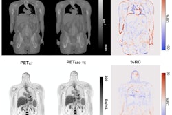
The dual-tracer protocol involves injecting patients first with C-11 acetate, followed 11 minutes later by scanning patients from the base of the skull to the upper thighs. Fifteen minutes after this, patients receive F-18 FDG radiotracer and then undergo whole-body imaging after 60 minutes.
Patients underwent conventional imaging for a median of 26 days prior to dual-tracer PET/CT. Of the entire cohort, 474 patients underwent CT, 46 underwent MRI, and five underwent both CT and MRI prior to dual-tracer PET/CT.
According to the findings, dual-tracer PET/CT had an accuracy of 95% for diagnosing HCC in indeterminate lesions on CT or MRI images. In addition, it identified new lesions in 14.3% of 273 staging patients and resulted in upstaging in 12% and treatment modifications in 7.7%.
Moreover, the technique upstaged 8.1% of 260 patients undergoing metastatic screening, the group reported.
“Dual-tracer PET/CT is routinely performed for HCC patients in our institution, and our findings are consistent with those of our prior studies and the reported literature,” the group wrote.
The authors noted that while the results are promising, dual-tracer PET/CT incurs a substantial radiation dose to patients, estimated to be around 29 mSv for men and 23 mSv for women. Comparatively, a CT scan of the abdomen typically delivers an effective dose of 10 mSV.
“However, we believe that the benefits of this imaging modality outweigh its potential risks, especially among the intended cohort of patients with high risks of cancer and reduced life expectancy,” the group concluded.
The full study is available here.