PET/MRI shows that young women with autism spectrum disorder (ASD) have increased brain metabolism associated with translocator protein (TSPO), a group from Harvard University in Boston has reported.
In a pilot study, the researchers showed that women with autism have higher uptake of carbon-11 (C-11) PBR28 radiotracer -- which binds to TSPO -- on brain PET/MRI than those without the condition. The finding adds to current scarce knowledge about molecular markers in these patients, noted lead author Chieh-En Jane Tseng, PhD, and colleagues.
“Over 90% of neuroimaging studies in ASD in the last 20 years only studied males or did not study sex effects if females were included. As a result, our understanding of neurobiology in females with ASD is particularly lacking,” the group wrote. The study was published April 13 in Neuropsychopharmacology.
Sex-based differences in the prevalence of ASD are well-documented, with a male-to-female ratio of approximately four to one, the authors explained. TSPO is a mitochondrial protein expressed in numerous cells including microglia, astrocytes, and neurons and is being investigated as a potential marker for neuroinflammation in certain diseases, including ASD.
Previous PET studies by the group suggest that men with ASD have a lower density of several mitochondrial proteins compared to healthy controls, yet to date, TSPO levels have not been investigated specifically in women with ASD, the researchers wrote.
To address the knowledge gap, the group enrolled 12 adult females with ASD and 10 age- and TSPO genotype-matched control patients to undergo PET/MRI imaging at the Lurie Center for Autism at Massachusetts General Hospital in Boston.
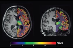
 Group mean C-11 PBR28 SUVR maps of females with ASD (top) and female controls (middle). Statistical map from voxelwise comparison of C-11 PBR28 SUVR between groups, controlled for age and TSPO genotype, shows elevated regional TSPO levels relative to whole brain mean in the midcingulate cortex and splenium of the corpus callosum in ASD (N = 12) compared with controls (N = 10) (Z > 2.3, pcluster) (bottom). TSPO =
translocator protein, ASD = autism spectrum disorder, CON = control, SUVR = standardized uptake value ratio, N = number. Image and caption courtesy of Neuropsychopharmacology.
The scans showed that women with ASD exhibited elevated C-11 PBR28 standardized uptake value ratios (SUVRs) in the midcingulate cortex and splenium of the corpus callosum compared to healthy controls. In addition, no brain area showed lower C-11 PBR28 SUVR in females with ASD compared with participants without ASD, according to the findings.
“Elevated regional C-11 PBR28 SUVR in females with ASD stand in stark contrast to our previous findings of lower regional C-11 PBR28 SUVR in males with ASD,” the researchers wrote.
Ultimately, such information is key to developing strategies and new approaches for treating ASD, the group wrote. Nonetheless, this was a small study, and additional research is warranted to determine whether changes in regional TSPO levels arise from the same process or cell source in males and females, they noted.
“Whether this contributes to sex differences in ASD prevalence and/or presentations of the core symptoms of ASD in individuals with ASD will need to be further elucidated,” the group concluded.
The full study is available here.