Radiographics 1997 Jan;17(1):63-79. Complex disease of the pleural space: radiographic and CT evaluation.
Kuhlman JE, Singha NK
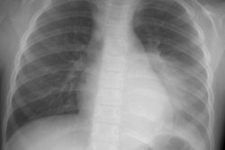
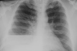
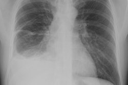
Pleural space disease is often complex, difficult to diagnose, and problematic to manage. At computed tomography (CT), empyema appears as an oblong fluid collection with smooth inner margins that compresses and displaces the surrounding lung and airways away from the pleural collection. CT findings in hemothorax include heterogeneous attenuation of pleural fluid, hyperattenuating areas of debris within pleural fluid, and a "fluid-hematocrit" level. Nodular pleural thickening at chest radiography or CT indicates a malignant pleural effusion; the cross-sectional capability of CT allows scrutiny of all pleural surfaces to detect enhancing tumor implants in addition to pleural effusions. Pleural plaque, rounded atelectasis, and pleural pseudotumor can mimic neoplastic disease on chest radiographs but can often be diagnosed with CT. Bronchopleural fistula may also be difficult to diagnose with radiography alone, necessitating further analysis with CT. Pleurocentesis fluid containing chyle, cerebrospinal fluid, amylase, or hyperalimentation fluid indicates pleural space disease with an unusual or iatrogenic cause. Recent advances in image-guided procedures have significantly improved treatment options for many complex pleural space processes.
PMID: 9017800, MUID: 97170271