Chylothorax:
Clinical:
Chyle is transported via the intestinal lymphatics to the cisterna chyli (which is located in the retrocrural space at approximately the level of the L1/L2 vertebral body) and then ascends in the thoracic duct. The thoracic duct courses through either the aortic or esophageal hiatus and ascends on the right side of the mediastinum between the azygous vein and the aorta. The duct crosses to the left at the level of T4-T8 and continues superiorly into the neck. Here the duct arches anteriorly over the subclavian artery and empties into the left subclavian or jugular vein. Up to 50% of individuals may have two ducts and there is considerable biologic variability.Chylothorax is the accumulation of lymphatic fluid in the pleural space and accounts for 2-3% of pleural effusions [4]. The diagnosis is confirmed with thoracentesis, which typically demonstrates milky pleural fluid with a triglyceride level greater than 110mg/dL (1.24 mmol/L) and a cholesterol level less than 200 mg/dL (5.18 mmol/L) [1,4].
Causes of chylothorax include:
- Trauma (25%) - The most common traumatic cause of thoracic duct disruption is surgery [1,4]. Esophageal surgery has the highest risk for traumatic chylothorax [4]. Injury to the thoracic duct is more often caused by penetrating trauma, than blunt trauma [2]. Hyperextension spine injury or posterior rib fracture can cause thoracic duct injury [2].
- Malignancy- this is the most common cause of non-traumatic chylothorax and lymphoma accounts for 70-75% of cases (lymphoma accounts for 39% of cases of chylothorax overall) [1,3,4]
- Congenital or idiopathic causes (15%).
Lymphangioleiomyomatosis accounts for under 10% of all cases of chylothorax, although up to 75% of patients affected with the disorder will have chylous effusions.
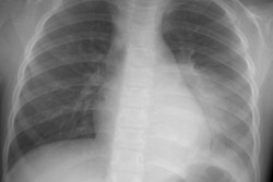
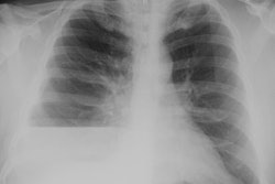
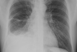
X-ray:
On CT, the fluid is usually not seen to be of fat density, because of its high protein content. Contrast material can be seen within the pleural space following lymphangiogram.REFERENCES:
(1) Radiographics 2002; Kim EA, et al. Radiographic and CT findings in complications following pulmonary resection. 22: 67-86
(2) AJR 2009; Raman SP, et al. Imaging of thoracic lymphatic
diseases. 193: 1504-1513
(3) Radiographics 2017; Bligh MP, et al. Spectrum of CT findings
in thoracic extranodal non-hodgkin lymphoma. 37: 439-461
(4) Radiographics 2020; Cholet C, et al. Nontraumatic
chylothorax: non-enhanced MR lymphography. 40: 1554-1573