J Comput Assist Tomogr 1997 Jan;21(1):115-120. Localized benign fibrous tumors of the pleura: MR appearance.
Ferretti GR, Chiles C, Cox JE, Choplin RH, Coulomb M
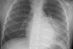
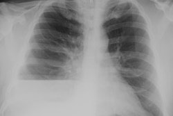
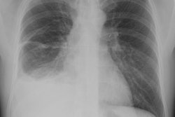
PURPOSE: Our goal is to describe the MR findings in benign localized fibrous tumors of the pleura. METHOD: Chest radiographs, CT scans, and MR images of four patients with localized benign fibrous tumors of the pleura were retrospectively reviewed and correlated with the pathologic findings. RESULTS: Tumors ranged from 4 to 18 cm in their largest diameter. Three tumors were located in the diaphragmatic region, and one was within the left major fissure. All tumors were round to ovoid, pedunculated, and well delineated. On T1-weighted SE MR images, tumors showed low signal intensity. All tumors had heterogeneous but predominantly low signal intensity on proton-density-weighted images and lower signal intensity on T2-weighted images. CONCLUSION: Localized benign fibrous tumors of the pleura were characterized by low signal intensity on all MR sequences that is explained by high collagen content within the tumors' stroma and should suggest the diagnosis preoperatively.
PMID: 9022782, MUID: 97175122