Primitive Neuroectodermal Tumor (Askin's tumor)
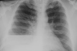
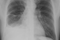
The CXR demonstrates a large left lung mass. The patient was initially felt to have a pneumonia, but subtle rib destruction can be seen involving the left posterior ninth rib. When the radiographic findings did not improve a CT scan was performed (See below). (Click images to enlarge)


CT demonstrated a large heterogeneous mass crossing the misline which contained areas of necrosis and calcification. There is posterior rib destruction, extension into the chest wall, and a pleural effusion.