CANCÚN, MEXICO - With the aid of a powerful transducer that can penetrate the skull, radiologists in Cuba have a versatile new ultrasound tool at their disposal for examining the head and neck.
At the 22nd International Congress of Radiology, Dr. Orlando Valls Pérez and Dr. Vivian Fleites Brague from Hospital Hermanos Ameijeiras in Havana, Cuba, discussed the head radiology applications of high-power ultrasound in a Spanish-language session Wednesday.
 |
| Dr. Orlando Valls Pérez. |
"Transcranial Doppler sonography is a new clinical and research application of ultrasound that allows a noninvasive examination to evaluate the brain circulation and cerebral perfusion, and measure blood-flow velocities in the cerebral vessels," Pérez said.
The system has multiple clinical applications in neurology, surgery, and stroke, as well as research, he said. The hospital uses a pulsed technique with 2-MHz pulse sound emission, very low frequency and high power, and a 7.5-MHz transducer with color-coded duplex Doppler. They use Combison 420 and Voluson 530D scanners manufactured by Kretz, Austria (now a GE Medical Systems company). Data can generally be obtained in the brain tissue anywhere between 25 mm and 100 mm from the transducer.
Transcranial ultrasound's advantages begin with noninvasiveness and low cost compared to other imaging methods. The patient can be examined without getting out of bed, and the system readily examines brain anatomy as well as the physiologic parameters of blood flow, according to Pérez. At the hospital, the technique has quickly evolved into a versatile real-time imaging tool.
Drawbacks include the difficulty of penetrating the very dense temporal bone, anatomical variations in the circle of Willis area, lesion asymmetry, and, like all ultrasound techniques, the need for a high level of operator skill and experience.
In neurology applications, the technique can be used to detect intracranial and extracranial stenosis, and to evaluate collateral circulation and vasomotor reactivity. It is routinely used for preoperative diagnoses of aneurysms and arteriovenous malformations, Pérez said, as well as in the detection of central nervous system tumors and brain necrosis by noting a lack of blood flow.
"Cerebrovascular diseases of the brain can be detected by increased or decreased blood flow, absence of the flow, and spectral changes," Pérez said. By comparing normal-velocity flows with measured flows, vessel patency can also be assessed. "Once you know the mean blood flow in a vessel, you have a very good idea of its physiology," he said.
 |
| Normal velocity values of intracranial blood-flow vessels. Chart courtesy of Dr. Orlando Valls Pérez. |
Surgical uses for the technique include diagnosis and follow-up of vasospasm, evaluation of head trauma, detection of emboli, and postoperative evaluation to prevent neurological complications. The technique has proven useful in interventional neuroradiology procedures and during cardiac surgery, Pérez said.
"It is very helpful for the surgeon because we can localize the tumor and carefully study its characteristics. We can see the relationship between the area of edema and normal tissues," Brague said.
Stroke applications include diagnosis of mid-cerebral artery occlusion, reperfusion follow-up, and the monitoring of thrombolytic therapy. In cerebral trauma cases, the technique has been used intraoperatively to detect emboli, as well as for clinical follow-up.
 |
| Depending on the region to be evaluated, transcranial Doppler ultrasound is performed by holding the transducer up to one of three windows in the head, including the orbital window (top) temporal window (left), and suboccipital window (bottom). Image courtesy of Dr. Orlando Valls Pérez. |
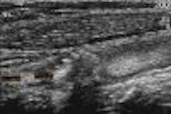
But the detection of aneurysms depends on their being large enough to visualize, generally be a centimeter or larger, and they must be located within the proximal segments of the circle of Willis arteries, Pérez said. They are characterized by the presence of a sacular image with a change in flow direction, and with low flow-velocity inside the aneurysm.
"Based on changes in the flow direction and mean velocity, we can then characterize (aneurysms) as mild, moderate, or severe," Pérez said.
 |
| Basal artery aneurysm in a 48-year-old male. Image courtesy of Dr. Orlando Valls Pérez. |
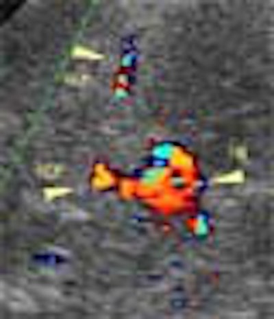
Arteriovenous malformations are evaluated by identifying "nests" of malformed blood vessels. They are characterized by increased systolic and diastolic blood flow with decreased hemodynamic resistance, as well as increased velocity and turbulence in the ipsilateral extracranial carotid artery, according to Brague.
"In this patient we can see the entire 'nest' of malformation, and identify in this case a very dilated mid-cerebral artery with fast flow and high diastolic values throughout," she said.
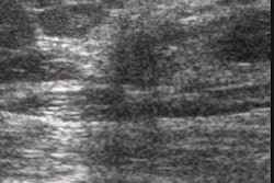
Brain tumors can be identified as echogenic masses surrounded by a hypoechoic halo. Heterogenous masses can be due to necrosis, cysts, or calcifications.
 |
| Cerebral teratoma in a 56-year-old male patient (left) with CT correlation (right). Images courtesy of Dr. Orlando Valls Pérez. |
"Intraoperatively, (transcranial Doppler sonography) guides the hand of the surgeon who is trying to separate normal from the abnormal tissue," during fine-needle aspirations and procedures to reduce cystic tumor volumes using aspiration techniques, Pérez said. "It helps to prevent hemorrhage and preserve more normal tissue."
By Eric Barnes
AuntMinnie.com staff writer
July 4, 2002
Copyright © 2002 AuntMinnie.com