A group of researchers has developed a new technique for elasticity ultrasound breast imaging that uses freehand scanning rather than mechanical compression. The group believes that the technique produces excellent images without patient discomfort and without the need for additional motorized components to be added to the scanner.
Dr. Timothy Hall and colleagues from the University of Kansas Medical Center in Kansas City discussed their preliminary research at the 2002 American Institute of Ultrasound in Medicine meeting in Nashville.
"A great deal of laboratory work has shown that images of the elastic properties of tissue can aid the diagnosis of breast lesions. However, these laboratory systems typically use motor-driven tissue compression, and this limits the flexibility of data acquisition," the group wrote in the program abstract.
To come up with a more practical application for clinical settings, they designed a software application that runs on a Sonoline Elegra premium scanner (Siemens Medical Solutions, Iselin, NJ). Additional external components are not required, they stressed. Motion was induced by pressing on the skin surface with a linear-array transducer and then tracked in 2-D using an adaptive block-matching algorithm.
 |
| A phantom with an 8-mm diameter spherical target that is three times stiffer than its background. The target is not visible in the standard B-mode ultrasound image (left), but is obvious in the strain image (right). |
"We describe our work with the term ‘palpation imaging’ because it more intuitively describes what we are doing," Hall wrote in an e-mail to AuntMinnie.com. "We press on the breast with the ultrasound transducer and watch the deformation in the typical ultrasound B-mode image, and the image of mechanical strain caused by that deformation, in real-time."
Tests were performed in phantoms with both motorized and freehand compression, while tests in volunteers were done with the freehand system, using a scanning technique similar to traditional breast sonography. The investigators addressed the issue of patient discomfort, which is a major drawback to mammography, according to many women.
"We warn each patient of the potential for discomfort prior to each scan," Hall said "We ask each patient during and after each scan about any discomfort, and there has been none. The total compression we attempt to achieve is significantly less than that of mammography, and it is momentary compared to the extended clamping used in mammography."
 |
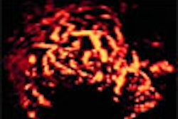
| The above image has some of the features typical of breast cancer: the tumor is hypoechoic (dark compared to its surroundings), and has an irregular boundary. The palpation image (an image of relative mechanical strain) of this lesion shows that the lesion is stiffer than its surroundings (dark compared to its background), and appears larger in the palpation image than in the B-mode image. |
According to the group’s results, freehand elasticity produces excellent image noise, contrast, and resolution compared to motorized compression. Elasticity image noise properties were slightly higher, while contrast and resolution were equivalent. The team found that small (2.4-mm diameter) spherical targets were easier to find with freehand scanning. They came up with similar results in the volunteer scans as well.
"We have had radiologists commenting on our image quality and on the information content of these images, and all comments are positive. The strain images in a compression/release sequence appear to be unique to specific lesion types. This is easily interpreted," Hall commented.
 |
| The B-mode image is not typical of cancer. It shows increased image brightness below the lesion, which is more characteristic of cysts. However, the lesion is very stiff compared to its surroundings, and considerably larger in the palpation image than in the B-mode image. This feature of relative size in the two imaging modalities is unique to cancers so far in this study. Both lesions were proven to be invasive ductal carcinomas after surgical excision. Images courtesy of Dr. Timothy Hall. |
While reading the images may not be an issue, this freehand technique will require additional training for sonographers in order to learn how to manipulate the scanner. Hall said that ultrasound technologists from his group were able to learn the technique quite rapidly, and were able to obtain acceptable images in 5-10 volunteer patients.
Hall said the group would continue to refine the software and perform experiments. The palpation imaging technique may be particularly useful for imaging women who cannot undergo traditional mammography, he added, as it has been successful in both young women with glandular breasts and older women with fatty breasts.
By Shalmali PalAuntMinnie.com staff writer
March 27, 2002
Related Reading
TechniScan unveils breast-specific ultrasound, February 13, 2002
Ultrasound has unique strengths in breast imaging, January 22, 2002
Ultrasound aids mammography in breast cancer diagnosis, December 3, 2001
New ultrasonic approach focuses on breast imaging, November 27, 2001
Copyright © 2001 AuntMinnie.com