Tissue harmonic imaging (THI) significantly improves the grayscale contrast between fatty tissue and focal lesions of the breast, particularly enhancing the conspicuity of lesions, according to ultrasound researchers.
Dr. Kazimierz Szopinski and colleagues presented their work at the 2002 American Institute of Ultrasound in Medicine meeting in Nashville, TN. Szopinski is from the department of diagnostic imaging at the Second Faculty of Medicine in Warsaw, Poland. His co-authors were based at the department of pathology at the Center of Medical Postgraduate Education in Warsaw and at the Centre d’Imagerie in Aix-en-Provence, France.
For the study, 69 focal lesions of the breast in 53 female patients were evaluated. There were 10 carcinomas, 53 benign solid lesions, and six benign cystic lesions. The women, with a mean age of 50.9 years, were all referred for fine-needle biopsy.
The lesions, which ranged in size from 3 mm to 10 cm, were imaged using THI and fundamental-frequency imaging on a Sonoline Elegra scanner (Siemens Medical Solutions, Iselin, NJ) with a wide-band linear-array 7.5-MHz transducer. The digital images were then transferred to a PC, where the grayscale intensity of the lesion, and adjacent adipose tissue, was measured using Adobe Photoshop software. The lesion-to-adipose tissue ration was calculated.
 |
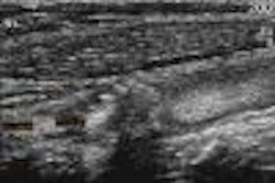
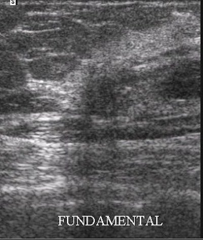
A 9-mm breast carcinoma. On the THI image, extensions of the tumor can be seen on the upper portion of the image, as well as in a very strong acoustic shadow beneath the tumor. There is clearly more grayscale contrast between the tumor and adjacent fatty tissue on the THI image. Images courtesy of Dr. Kazimierz Szopinski.
 |
According to the results, the mean grayscale contrast between the lesions and the fatty tissue was 0.38 in the fundamental frequency and 0.51 on THI. In 88.4% (61) of the cases, the contrast between the lesion and the fatty tissue was better in the THI images, and the contrast improvement reached statistical significance (P<.001), they reported.
"We believe THI would be particularly beneficial in predominantly fatty breasts, (such as) obese and/or elderly patients," Szopinski wrote in an e-mail to AuntMinnie.com.
While other researchers have found that THI can reduce scan time, Szopinski said his group did not find that to be the case. However, "THI is a problem-solving tool when a lesion has echogenicity close to the echnogenicity of the fatty breast tissue. THI also enhances the acoustic shadows, which are an important sonographic sign."
Szopinski said he does not use THI for the first-pass scan in his practice, but relies on the technique when studying particular lesions or suspicious areas of the breast.
By Shalmali PalAuntMinnie.com staff writer
May 10, 2002
Related Reading
Sonographers quantify gains with tissue harmonic imaging, April 3, 2000
Breast lesion classification may improve with US technology, January 5, 2000
Copyright © 2002 AuntMinnie.com