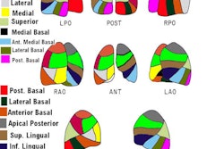
Modified PIOPED II Criteria (From: J Nucl Med 2008; Sostman HD, et al. Sensitivity and specificity of perfusion scintigraphy combined with chest radiography for acute pulmonary embolism in PIOPED II. 49: 1741-1748)
| PE Present | High probability (2 or more segments of perfusion-chest radiograph mismatch) |
| PE Absent | Normal perfusion |
| Very low probability
- Nonsegmental lesion (prominent hilum, cardiomegaly, elevated diaphragm, linear atelectasis, of CP angle effusion) with no other perfusion defect in either lung) - Perfusion defect smaller than radiographic lesion - 1-3 small segmental defects - Solitary chest radiograph-perfusion matched defect in mid- or upper lung zone confined to a single segment - Stripe-sign around perfusion defect (best tangential view) - Pleural effusion in at least one-third of pleural cavity, with no other perfusion defect in either lung |
|
| Not diagnostic | All other findings |
Trinary Interpretation System (From J Nucl Med 2011; Glasser JE, et al. Successful and safe implementation of a trinary interpretation and reporting strategy for V/Q lung scinitgraphy. 52: 1508-1512
| PE Positive | High probability (a single or more segmental
mismatch) |
| PE Negative |
Normal perfusion, very low, or low probability
scans |
| Not diagnostic | Intermediate or indeterminate exams |