CHICAGO -- AI-assisted diffusion-weighted imaging (DWI) shows differences in the brains of children with and without attention-deficit/hyperactivity disorder (ADHD), a study presented November 29 at the 2023 RSNA annual meeting has found.
In his presentation, Justin Huynh from the University of California, San Francisco found that the deep-learning model revealed significant differences in nine brain white matter tracts in children with ADHD.
“These differences in MRI signatures in individuals with ADHD have never been seen before at this level of detail,” Huynh said. “In general, the abnormalities seen in the nine white matter tracts coincide with the symptoms of ADHD.”
Reports estimate that 5.7 million U.S. children and adolescents between the ages of 6 and 17 years have been diagnosed with ADHD, which can continue into adulthood. Early diagnosis and intervention strategies help manage this condition.
Huynh noted that diagnosing ADHD is complex and relies on criteria that are susceptible to subjective biases. He said that imaging could provide abundant data on the underlying psychopathology and etiology of this condition. However, Huynh added that the variations seen in imaging between children with and without ADHD aren’t well understood.
Huynh and colleagues used DWI with unsupervised deep learning to visualize derived white matter tractography and fractional anisotropy measurements among adolescent patients with ADHD.
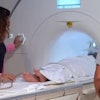
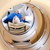
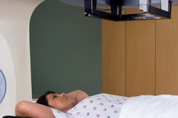
 Child undergoing pediatric MRI. Image and caption courtesy of RadiologyInfo.org.
The researchers used DWI data from a subset of 1,704 pediatric patients from the Adolescent Brain Cognitive Development (ABCD) Study. After training the deep learning model, the team evaluated it on an independent testing set of 333 patients. Of these, 193 had a behavior problem monitor (BPM) score greater than 70, the clinical threshold for ADHD. The other 140 had a BPM of less than 60, meaning no ADHD.
The team found that the average absolute error between predicted and actual fractional anisotropy values was 0.041. It reported that this was significantly different between subjects with (0.042) and without ADHD (0.038, p = 0.041).
The researchers also compared the absolute error anomaly scores between children with and without ADHD along 30 white matter tracts and found significant differences in the following nine tracts: left arcuate fasciculus, left cingulum cingulate gyrus, left corticospinal tract, right corticospinal tract, frontoparietal, right inferior fronto-occipital fasciculus, left inferior longitudinal fasciculus, superior parietal lobe, and temporal lobe.
In these tracts, the fractional anisotropy was significantly elevated in children with ADHD. All achieved p-values of less than 0.05 when compared with imaging data from children without ADHD.
Huynh highlighted that this method provides a promising step toward finding imaging biomarkers that can be used to diagnose ADHD in a quantitative, objective diagnostic framework.
Huynh said the team will continue gathering data from the other participants in the ABCD study and compare the performance of additional AI models.