A group in Boston has validated an AI algorithm that could be useful for identifying nonsmokers who are at high risk of lung cancer, according to a study to be presented at RSNA 2023 in Chicago.
A team led by medical student Anika Walia, of Boston University, who will present the study, developed a model called CXR-Lung-Risk using 147,497 chest x-rays of 40,643 asymptomatic smokers and never-smokers from a previous lung cancer screening trial.
“Using routine CXRs from the [electronic medical records], CXR-Lung-Risk identified never-smokers at high risk of lung cancer, a group in which lung cancer rates are increasing,” Walia said, in a news release from RSNA.
The American Cancer Society estimates about 238,340 new cases of lung cancer will be diagnosed in the U.S. this year, with up to 20% of these cases occurring in "never-smokers" – people who have never smoked or smoked fewer than 100 cigarettes in their lifetime.
In this study, the researchers externally validated the model in a separate group of never-smokers who underwent routine outpatient chest x-rays from 2013 to 2014. The primary outcome was six-year incident lung cancer, identified using International Classification of Disease codes. Risk scores were then converted to low, moderate, and high-risk groups based on externally derived risk thresholds.
Of 17,407 patients (mean age 63 years) included in the study, 28% were deemed high risk by the deep learning model, and 2.9% of these patients later had a diagnosis of lung cancer, according to the findings. In addition, the high-risk group well exceeded the 1.3% six-year risk threshold where lung cancer screening CT is recommended by National Comprehensive Cancer Network (NCCN) guidelines.
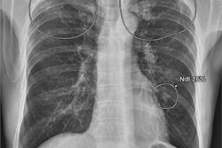
 An example of a patient frontal chest x-ray showing a subtle nodular opacity (arrow) identified by AI in the right middle lung zone. Axial, noncontrast, low-dose chest CT scan shows a 1.1-cm solid nodule (arrow) in the right lower lobe. Image courtesy of Anika Walia, Boston University
Moreover, after adjusting for age, sex, race, previous lower respiratory tract infection, and prevalent chronic obstructive pulmonary disease, there was still a 2.1 times greater risk of developing lung cancer in the high-risk group compared with the low-risk group.
“A major advantage to our approach is that it only requires a single chest x-ray image, which is one of the most common tests in medicine and widely available in the electronic medical record,” Walia noted.
Ultimately, the U.S. Preventive Services Task Force (USPSTF) currently recommends lung cancer screening with low-dose CT for adults between the ages of 50 and 80 who have at least a 20 pack-year smoking history and currently smoke or have quit within the past 15 years. The USPSTF does not recommend screening for individuals who have never smoked or who have smoked very little, however.
With the incidence of lung cancer among never-smokers on the rise, the CXR-Lung-Risk model could potentially serve as a tool for early detection through screening in these cases, the researchers suggested.
“Since cigarette smoking rates are declining, approaches to detect lung cancer early in those who do not smoke are going to be increasingly important,” added senior author Michael Lu, MD, of Harvard Medical School.