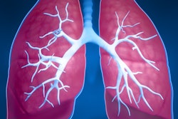
Examining several radiologic features of lung nodules on CT scans enabled radiologists to discern whether the nodules were benign or cancerous, according to an article published online May 14 in PLOS One.
Looking to lower the rate of false positives in CT lung cancer screening, researchers from the Mayo Clinic in Rochester, MN, and Vanderbilt University reviewed a selection of 318 benign and 408 malignant lung nodules (> 7 mm) on the CT scans of participants in the National Lung Screening Trial (NLST). They tested the ability of 57 independent quantitative variables to characterize the nodules based on radiologic features such as volume, shape, and texture.
Among these variables, they identified eight that were able to distinguish between benign and malignant nodules with statistically significant accuracy (p < 0.01): vertical location, volume estimate, flatness, texture analysis, maximum shape index, average shape index, average positive mean curvature, and minimum mean curvature. The area under the curve for these eight features was 0.939.
The radiomics approach looks promising and has the potential to change the way physicians evaluate incidentally detected lung nodules, said first author Dr. Tobias Peikert in a statement from the Mayo Clinic.