The use of 3D-printed heart models in healthcare has improved the management of patients with congenital heart disease (CHD) on multiple fronts, from surgical planning and simulation to patient education, according to a review published online April 30 in JACC: Basic to Translational Science.
The anatomical complexity and high-risk surgery involved with CHD care make it a prime candidate for the application of 3D printing, lead author Dr. Shafkat Anwar, a pediatric cardiologist at Washington University School of Medicine in St. Louis, told AuntMinnie.com (JACC: Basic to Transl Sci, April 30, 2018).
"CHD interventions are high-risk procedures, and presurgical planning requires the mental reconstruction of complex 3D anatomic information," he said. "3D printing provides a replica of the patient's anatomy ... [and] using these models enables precise presurgical planning and simulation. This will hopefully improve patient outcomes."
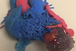
 Multicolor 3D-printed heart. All images courtesy of Dr. Shafkat Anwar.
Though the adoption of 3D printing in cardiology is relatively new, clinicians have already reported a wide range of uses and benefits associated with the technology, including the following:
- Patient education
- Presurgical planning
- Intraoperative guidance
- Medical device and implant production
Several groups have reported success in planning and simulating the repair of damaged heart valves, ventricles, and vessels requiring complex procedures such as the Fontan procedure and intracardiac baffle repair, according to the authors. More than simply allowing surgeons to prepare for a single task, 3D-printed hearts have provided them with a means of simulating "plan B" and "bailout" scenarios for high-risk surgeries and developing patient-tailored surgical plans -- neither of which are feasible with the current standard of care.
Clinicians commonly use solid, or "blood-pool," 3D-printed hearts to plan such procedures and "hollow" models for simulation, the authors noted. Recently, surgeons have been simulating surgeries using 3D-printed hearts composed of flexible material to enhance realism.
What's more, 3D printing technology could help lower the learning curve for challenging cases and engender a shift in training from the traditional apprenticeship model to a simulator-based learning experience. 3D printing could also promote multidisciplinary communication -- encouraging physicians from different specialties to discuss pathology, likely outcomes, and perioperative care, which could reduce medical errors, Anwar said.
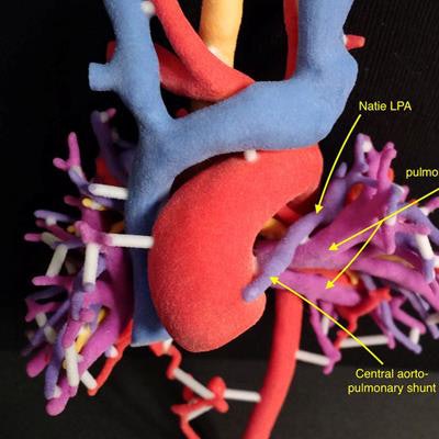
 3D-printed pulmonary and aortic vasculature.
What does the future entail for 3D printing and congenital heart disease?
Anwar named three key ways in which advances in 3D printing may ultimately improve care for patients:
- Expanded 3D visualization: 3D printing can complement advanced visualization technologies and potentially allow surgeons to manipulate electronic 3D models in real-time using augmented or virtual reality.
- Integration of physiology with anatomy: "Integration of 4D flow and computational fluid dynamics will enable more accurate simulations of postoperative anatomy and physiology," he said.
- Advanced materials: "Advances in tissue-mimicking materials will lead to more realistic models, and bioprinting may be a game-changer," he said.
Anwar's future research interests include the study of outcomes related to 3D printing and integration of multiple imaging modalities, such as 3D echocardiography, CT angiography, and MRI, with 3D printing.