PEBBLE BEACH, CA - Radiography may be the first choice for assessing the painful hip, but MRI techniques offer an opportunity to perform customized imaging, which is especially important if invasive surgery is in a patient's future, according to a presentation given Thursday at the Orthopedic Imaging at Pebble Beach conference.
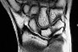
Dr. Robert Boutin, the chief of musculoskeletal imaging and the medical director of Franklin, TN-based MedTel International, started his talk by reviewing the basic anatomy of the ball-and-socket setup that composes the hip joint. In terms of osseous anatomy, the acetabulum and the femur are particularly important sites as MR is relied upon to assess abnormalities. The cartilage and labrum make up the hip joint's articular anatomy, while extrinsic and intrinsic ligaments hold the joint capsule together.
"All of these are very important anatomic structures. They all may be operated on these days," said Boutin, who is based in Davis, CA. "We really have to tailor our MRI techniques to look for internal derangements in these structures if the patient is a potential operative candidate."
MRI protocols and disease pathology
Boutin described three major MR protocols that apply to patients who are sent for hip imaging: one for routine screening, one to assess for internal derangement, and one to reduce artifacts in patients with prosthetic hardware.
For the clinical indication of avascular necrosis (AVN) in a screening setting, Boutin recommended a large field-of-view (FOV, 46-40 cm) to screen both hips in the coronal and axial planes, while using a smaller FOV to take a closer look at the affected hip in the sagittal plane (≤ 20-40 cm).
The causes of AVN can either be traumatic injury (hip subluxation) or a nontraumatic systemic problem, such as thromboembolic abnormalities. Those with systemic AVN are at risk for osteonecrosis in several other sites as well, Boutin said.
The sensitivity, specificity, and accuracy of MRI in AVN has held fast at 90% or more in several studies, he noted, adding that many difference sequences can be used for screening. He cited a protocol of coronal T1-weighted and fast spin-echo (FSE), axial T2-weighted and FSE, and sagittal T1-weighted as an example of one that is often used.
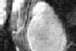
Common MRI findings for AVN include a demarcated zone of altered signal intensity extending to the superior subchondral bone plate, a double-line sign on T2-weighted images, and a serpentine border on low signal intensity images.
When reporting to orthopedic specialists, Boutin said the most important hallmarks of AVN to relate are femoral head collapse, size of AVN, and acetabular changes.
Moving on to the assessment of internal derangement, Boutin said that high-resolution, unilateral hip imaging is a must for diagnostic success. He recommended using a surface coil along with a small FOV (14-16 cm), section thickness of 3-4 mm, and a "negligible interslice gap."
"Just like we would never think of looking for a meniscus tear in the knee by scanning both knees at the same time, if we are looking for an internal derangement of the hip, we shouldn't be scanning both hips at the same time," he said.
MR arthography (MRA), with an average of 10 mL of injected gadolinium (off-label use), is another option and is generally preferred by radiologists for assessing the hip labrum and articular cartilage, Boutin said. MRA does require STIR images to assess extra-articular abnormalities that may not be seen on T1-weighted imaging, he explained.
"The added benefit of MRA is that when you have the needle in that joint, you can concurrently inject local anesthetic and confirm that the pain generator is in the hip," Boutin said. "If the pain goes away after injection of the local anesthetic, the presumption is that the pain generator is in the hip. If the pain doesn't go away, then you really have to consider the possibility of pain being referred to the hip region from another place such as the spine."
Among the various causes of labral derangements is developmental dysplasia of the hip (DDH) and femoroacetabular impingement (FAI). FAI generally strikes people under the age of 40 and is considered a major cause of osteoarthritis, Boutin explained. There are two types of FAI, femoral FAI (also known as cam FAI) and acetabular FAI (pincer FAI), and most cases are a combination of both.
On MRI, the main criterion for a labral tear is a linear hyperintense signal contacting the labral surface, Boutin wrote in the course syllabus. This signal can appear either at the labral-acetabular junction or the labrum itself. If the labra appears with enlarged or blurred margins, or demonstrates indeterminate signal intensity, then degeneration has most likely occurred.
"It's helpful to know where labral tears and derangement occur," Boutin said. "Commonly, they occur in the anterior or anterosuperior portion of the acetabular labrum. Coronal images commonly don't show the tear. Axial images often don't show the tear. Oblique axial images should show the tears with an 88% sensitivity. This is the best way to see these labral detachments."
MRA is currently considered more accurate than standard MRI for diagnosing labral tears, but detection depends on the size of the labral lesion. Also, a serious pitfall to watch out for on MRI is the misdiagnosis of sublabral sulcus as a tear. On MRI, a sublabral sulcus will manifest with smooth edges and the absence of adjacent degenerative changes.
Finally, there is a protocol for patients with metal implants, a situation that was once considered a contraindication for MR exams, Boutin said. However, MRI has proved to be most suitable for investigating the causes of pain after total hip arthroplasty, such as a loose component, stress fracture, or nerve compression.
Boutin recommend the following low-field MR protocol for reducing metallic susceptibility artifacts: FSE imaging with minimal interecho spacing, STIR sequence, and no gradient-recalled echo (GRE) sequence.
He also outlined a newer protocol called MARS, or metal artifact reduction series, that advocates the following adjustments: increase receiver bandwith and number of excitations, increase spatial resolution, and direct misregistration artifacts away from the areas of diagnostic interest.
With misregistration artifacts, "you have control over what is called the frequency encoding direction," Boutin explained. "It turns out that the misregistration artifacts preferentially occur in this frequency encoding direction. For example, if you want to see medial and lateral to an implant, you can direct the frequency coding direction in an inferior to superior direction."
Other hip disorders
Boutin offered some tips on what to look for on MRI when the following conditions are suspected:
Transient bone marrow edema: Ill-defined low T1-weighted and high T2-weighted signal intensity in the femoral head and neck. No double line sign.
Fractures: The majority of young patients with femoral neck stress fractures will show resolution of signal intensity abnormalities on inversion-recovery MR sequences within six months. Persistent signal intensity indicates residual or recurrent injury. In intertrochanteric fractures, MRI can make the definitive diagnosis as to whether the fracture crosses the midline.
Arthritis: An optimized fat-suppressed spoiled gradient-recalled (SPGR) sequence with an 8 cm FOV is best for depicting articular cartilage lesions. Cartilage loss of less than 1 cm is not seen very well on MRI or MRA. In inflammatory arthritis, MRI can demonstrate joint effusion. In septic arthritis, use MRI for finding joint effusion with internal debris and marginal erosions.
By Shalmali Pal
AuntMinnie.com staff writer
February 2, 2007
Related Reading
Dietary changes might curb skeletal dysplasia, January 1, 2007
Low cytochrome P450 activity may trigger steroid-induced osteonecrosis, November 26, 2006
Multidetector CT better than MRI for assessing hip articular cartilage, August 17, 2005
Copyright © 2007 AuntMinnie.com