Using 4D flow MRI to show abnormal systolic flows in the aortas of patients with a bicuspid aortic valve, researchers in California hope to identify patients at risk of developing ascending aortic aneurysm.
The study, published in the April issue of Radiology, was conducted at the University of California, San Francisco (UCSF) and Stanford University School of Medicine in Stanford, CA. The lead author is Dr. Michael Hope from UCSF (Radiology, April 2010, Vol. 255:1, pp. 53-61).
Previous studies have shown that bicuspid aortic valve abnormalities may account for more morbidity and mortality than all other congenital cardiac malformations combined. Now, with the help of time-resolved 3D phase-contrast MRI -- also know as 4D flow MRI -- researchers can evaluate multidirectional blood-flow velocity data in the thoracic aorta.
Patient sample
4D flow MRI was used to assess blood flow in the thoracic aorta of 53 subjects -- 20 patients with a bicuspid aortic value, 25 patients with a tricuspid aortic valve, and eight healthy volunteers.
MRI scans were performed on a 1.5-tesla system (Signa CV/i, GE Healthcare, Chalfont St. Giles, U.K.) with an eight-channel cardiac coil, respiratory compensation, and retrospective electrocardiographic gating. 4D flow MRI was performed after standard cardiac MRI in all patients.
4D flow MRI uses a radiofrequency-spoiled gradient-echo pulse sequence (repetition time/echo time, 4.6-5.0 msec/1.7-2.0 msec; flip angle at 15°; velocity encoding, 160-200 cm/sec; fractional field-of-view, 300 x 270 mm; slab thickness, 78 mm; matrix, 256 x 192 x 30; spatial resolution, 1.17 x 1.56 x 2.60 mm; temporal resolution, 74-77 msec) and an oblique-sagittal slab encompassing the thoracic aorta.
The researchers used a total of 735 heartbeats for data acquisition, resulting in imaging times of eight to 15 minutes, with a mean time of 11 minutes.
 |
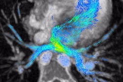
| Images show normal systolic flow in a patient with a tricuspid aortic valve (TAV) and normal thoracic aorta dimensions. Above, 4D flow MRI data in an oblique-sagittal orientation with 3D streamlines (color coded for velocity) during peak systole. Left: from right side of thoracic aorta. Right: from left side of thoracic aorta. Note smooth trajectory and absence of substantial secondary flow features. Below, close-up of area in white box in first image. Bottom image: Vector analysis at sinotubular junction during peak systole. Left: cross-sectional depiction. Right: sagittal depiction. Note relatively central velocity profile. Images © Radiology, April 2010, Vol. 255:1, pp. 53-61, Radiological Society of North America. |
 |
 |
The 4D flow MRI scans revealed nested helical flow at peak systole in the ascending aorta of 15 of 20 patients with a bicuspid aortic valve. However, no nested helical flow was detected in any of the healthy volunteers or patients with a tricuspid aortic valve.
The nested helical flow pattern was evident in six patients with a bicuspid aortic valve with a dilated ascending aorta and in nine patients with a normal ascending aorta. It also was seen in nine patients with no aortic stenosis.
In patients with a normal ascending aorta diameter, 4D flow MRI's evaluation during peak systole revealed no secondary flow features in the eight healthy volunteers, or in 21 patients with a tricuspid aortic valve and normal aortic dimensions.
4D MR images
The 4D flow MRI scans also showed that 10 of 11 patients with dilated ascending thoracic aorta also had abnormal secondary flow features. In addition, four patients with a tricuspid aortic valve with dilated ascending aortas had an abnormal systolic flow pattern, while six of seven patients with a bicuspid aortic valve with dilated ascending aortas had marked right-handed nested helical systolic flow.
Of the 13 patients with a bicuspid aortic valve and normal aortic dimensions, four patients had normal flow and nine patients had nested helical systolic flow. Five of those nine patients with nested helical flow were younger than 20 years of age, ranging from 6 to 19 years, while the other four patients were between 30 and 45 years old.
Based on the findings, the researchers concluded that 4D flow MRI proves that "markedly abnormal helical flow can be seen in the ascending thoracic aorta of patients with a bicuspid aortic valve, including those without aneurysm or aortic stenosis. Identification and characterization of eccentric flow jets in these patients may help identify those at risk for development of an ascending aortic aneurysm."
The authors cited several limitations of the study, including the lack of follow-up on the patients with a bicuspid aortic valve who had abnormal systolic flow in the ascending aorta.
"This was beyond the scope of our study," the authors wrote, "which was aimed at identifying and characterizing abnormal flow patterns in the context of a bicuspid aortic valve. To further evaluate the proposed hypothesis that eccentric flow jets are implicated in ascending aortic dilation, data documenting increased rates of growth are needed."
By Wayne Forrest
AuntMinnie.com staff writer
April 5, 2010
Related Reading
MRI shows age- and gender-based differences in myocardial motion, December 18, 2009
4D MRI enables blood-velocity measurement in mitral valve insufficiency, June 24, 2005
Copyright © 2010 AuntMinnie.com