Researchers have made available a collection of 304 T1-weighted MR images so that their counterparts worldwide can develop algorithms to better evaluate stroke patients and personalize treatment. The international group described the work in a study published online February 20 in Scientific Data.
The open-source dataset is known as the Anatomical Tracings of Lesions After Stroke (ATLAS). Through this resource of manually segmented lesions and metadata, the researchers hope to identify biological markers that could predict which rehabilitation therapies will produce the best response.
"One of our goals is to meta-analyze thousands of stroke MRIs from around the world to understand how the lesions impact recovery," said lead author Sook-Lei Liew, PhD, an assistant professor at the University of Southern California (USC), in a release from the university. "We are really interested in helping find better automated ways, using machine learning and computer vision, to identify the lesions and have machines draw those boundaries."
ATLAS data are stored by the International Neuroimaging Data-Sharing Initiative (INDI) and are available for download. Thus far, 33 research groups from Finland, Iran, Australia, and other countries are using the segmented MR images to create and validate algorithms that can automatically process results from stroke patients.
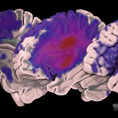
 Images depict the distribution of lesions in the dataset: the redder the color, the greater the number of patients with lesions in that part of the brain. Courtesy of USC Mark and Mary Stevens Neuroimaging and Informatics Institute/Tyler Ard.
Typically, researchers manually draw boundaries around stroke-related lesions. With the help of new algorithms, an automated segmentation process would be able to analyze more images.
"Ultimately, we would run their data through an automated pipeline that would give us some measures of [patients'] likelihood of recovery, or more importantly, their likelihood of responding to different types of therapies," Liew said. "We could then personalize their rehabilitation therapy based on their MRI results and, hopefully, improve their recovery."
Stroke researchers who wish to access the data can download a normalized subset from the INDI or the full dataset from the Inter-University Consortium for Political and Social Research.